 Movie
Movie Controller

[English] 日本語
 Yorodumi
- PDB-1icp: CRYSTAL STRUCTURE OF 12-OXOPHYTODIENOATE REDUCTASE 1 FROM TOMATO ... -
+ Open data

- Basic information
| Entry | Database: PDB / ID: 1icp | ||||||
|---|---|---|---|---|---|---|---|
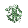
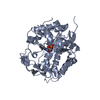
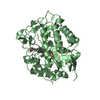
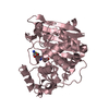
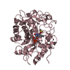
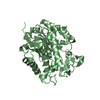
| Title | CRYSTAL STRUCTURE OF 12-OXOPHYTODIENOATE REDUCTASE 1 FROM TOMATO COMPLEXED WITH PEG400 | ||||||
 Components Components | 12-OXOPHYTODIENOATE REDUCTASE 1 | ||||||
 Keywords Keywords | OXIDOREDUCTASE / beta-alpha-barrel / PROTEIN-FMN-PEG complex | ||||||
| Function / homology |  Function and homology information Function and homology information12-oxophytodienoate reductase / 12-oxophytodienoate reductase activity / oxylipin biosynthetic process / fatty acid biosynthetic process / FMN binding / oxidoreductase activity / cytoplasm Similarity search - Function | ||||||
| Biological species |  | ||||||
| Method |  X-RAY DIFFRACTION / SYNCHROTRON / MOLECULAR REPLACEMENT / Resolution: 1.9 Å X-RAY DIFFRACTION / SYNCHROTRON / MOLECULAR REPLACEMENT / Resolution: 1.9 Å | ||||||
 Authors Authors | Breithaupt, C. / Strassner, J. / Breitinger, U. / Huber, R. / Macheroux, P. / Schaller, A. / Clausen, T. | ||||||
 Citation Citation | Journal: Structure / Year: 2001 Title: X-ray structure of 12-oxophytodienoate reductase 1 provides structural insight into substrate binding and specificity within the family of OYE. Authors: Breithaupt, C. / Strassner, J. / Breitinger, U. / Huber, R. / Macheroux, P. / Schaller, A. / Clausen, T. | ||||||
| History |
| ||||||
| Remark 600 | HETEROGEN THE LIGAND 2PE IS ALSO REFERRED TO AS PEG400. |
- Structure visualization
| Structure viewer | Molecule: MolmilJmol/JSmol |
|---|
- Downloads & links
-Download
| PDBx/mmCIF format | 1icp.cif.gz | 165.5 KB | Display | PDBx/mmCIF format |
|---|---|---|---|---|
| PDB format | pdb1icp.ent.gz | 127.9 KB | Display | PDB format |
| PDBx/mmJSON format | 1icp.json.gz | Tree view | PDBx/mmJSON format | |
| Others |  Other downloads Other downloads |
-Validation report
| Arichive directory | https://data.pdbj.org/pub/pdb/validation_reports/ic/1icpftp://data.pdbj.org/pub/pdb/validation_reports/ic/1icp | HTTPS FTP |
|---|
-Related structure data
| Related structure data |  1icqC  1icsC  1oyaS C: citing same article ( S: Starting model for refinement |
|---|---|
| Similar structure data |


-Links
 PDBj
PDBj- Assembly
| Deposited unit | 
| ||||||||
|---|---|---|---|---|---|---|---|---|---|
| 1 | 
| ||||||||
| 2 | 
| ||||||||
| Unit cell |
|
-Components
-Protein , 1 types, 2 molecules AB
| #1: Protein | Mass: 42443.035 Da / Num. of mol.: 2 / Mutation: R142M Source method: isolated from a genetically manipulated source Source: (gene. exp.)  |
|---|
-Non-polymers , 5 types, 490 molecules 


| #2: Chemical | ChemComp-CL /  Mass: 35.453 Da / Num. of mol.: 1 / Source method: obtained synthetically / Formula: Cl Mass: 35.453 Da / Num. of mol.: 1 / Source method: obtained synthetically / Formula: Cl | ||||||
|---|---|---|---|---|---|---|---|
| #3: Chemical |  Mass: 96.063 Da / Num. of mol.: 2 / Source method: obtained synthetically / Formula: SO4 Mass: 96.063 Da / Num. of mol.: 2 / Source method: obtained synthetically / Formula: SO4#4: Chemical |  Mass: 456.344 Da / Num. of mol.: 2 / Source method: obtained synthetically / Formula: C17H21N4O9P Mass: 456.344 Da / Num. of mol.: 2 / Source method: obtained synthetically / Formula: C17H21N4O9P#5: Chemical |  Mass: 414.488 Da / Num. of mol.: 2 / Source method: obtained synthetically / Formula: C18H38O10 / Comment: precipitant*YM Mass: 414.488 Da / Num. of mol.: 2 / Source method: obtained synthetically / Formula: C18H38O10 / Comment: precipitant*YM#6: Water | ChemComp-HOH / | Mass: 18.015 Da / Num. of mol.: 483 / Source method: isolated from a natural source / Formula: H2O |
-Experimental details
-Experiment
| Experiment | Method: X-RAY DIFFRACTION / Number of used crystals: 1 |
|---|
- Sample preparation
| Crystal | Density Matthews: 2.81 Å3/Da / Density % sol: 56.16 % | ||||||||||||||||||||||||||||||
|---|---|---|---|---|---|---|---|---|---|---|---|---|---|---|---|---|---|---|---|---|---|---|---|---|---|---|---|---|---|---|---|
| Crystal grow | Temperature: 291 K / Method: vapor diffusion, sitting drop / pH: 7.5 Details: Tris/HCl, ammonium sulfate, PEG400, pH 7.5, VAPOR DIFFUSION, SITTING DROP, temperature 291K | ||||||||||||||||||||||||||||||
| Crystal grow | *PLUS Temperature: 18 ℃ / pH: 7 | ||||||||||||||||||||||||||||||
| Components of the solutions | *PLUS
|
-Data collection
| Diffraction | Mean temperature: 100 K |
|---|---|
| Diffraction source | Source: SYNCHROTRON / Site: EMBL/DESY, HAMBURG  / Beamline: BW7A / Wavelength: 1.033 Å / Beamline: BW7A / Wavelength: 1.033 Å |
| Detector | Type: MARRESEARCH / Detector: CCD / Date: Nov 28, 1999 |
| Radiation | Protocol: SINGLE WAVELENGTH / Monochromatic (M) / Laue (L): M / Scattering type: x-ray |
| Radiation wavelength | Wavelength: 1.033 Å / Relative weight: 1 |
| Reflection | Resolution: 1.9→25 Å / Num. obs: 66928 / % possible obs: 91.8 % / Redundancy: 1.7 % / Biso Wilson estimate: 16.1 Å2 / Rmerge(I) obs: 0.049 / Net I/σ(I): 13.1 |
| Reflection shell | Resolution: 1.9→1.96 Å / Redundancy: 1.6 % / Rmerge(I) obs: 0.343 / Mean I/σ(I) obs: 1.9 / Num. unique all: 5356 / % possible all: 88.5 |
| Reflection | *PLUS Num. measured all: 101396 |
| Reflection shell | *PLUS % possible obs: 88.5 % |
- Processing
| Software |
| |||||||||||||||||||||||||
|---|---|---|---|---|---|---|---|---|---|---|---|---|---|---|---|---|---|---|---|---|---|---|---|---|---|---|
| Refinement | Method to determine structure: MOLECULAR REPLACEMENT Starting model: pdb: 1OYA Resolution: 1.9→22.9 Å / Cross valid method: THROUGHOUT / Stereochemistry target values: Engh & Huber
| |||||||||||||||||||||||||
| Displacement parameters | Biso mean: 26 Å2 | |||||||||||||||||||||||||
| Refinement step | Cycle: LAST / Resolution: 1.9→22.9 Å
| |||||||||||||||||||||||||
| Refine LS restraints |
| |||||||||||||||||||||||||
| Software | *PLUS Name: CNS / Version: 1 / Classification: refinement | |||||||||||||||||||||||||
| Refinement | *PLUS Highest resolution: 1.9 Å / Lowest resolution: 22.9 Å / % reflection Rfree: 5 % / Rfactor obs: 0.182 | |||||||||||||||||||||||||
| Solvent computation | *PLUS | |||||||||||||||||||||||||
| Displacement parameters | *PLUS Biso mean: 26 Å2 | |||||||||||||||||||||||||
| Refine LS restraints | *PLUS
|
