 Movie
Movie Controller


+ Open data

- Basic information
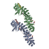
| Entry | Database: PDB / ID: 1i7x | ||||||
|---|---|---|---|---|---|---|---|
| Title | BETA-CATENIN/E-CADHERIN COMPLEX | ||||||
 Components Components |
| ||||||
 Keywords Keywords | CELL ADHESION / E-cadherin / beta-catenin / protein-protein complex / extended interface / armadillo repeat | ||||||
| Function / homology |  Function and homology information Function and homology informationlung cell differentiation / epicardium-derived cardiac vascular smooth muscle cell differentiation / mesenchyme morphogenesis / RUNX3 regulates WNT signaling / Regulation of CDH11 function / cardiac vascular smooth muscle cell differentiation / Regulation of MITF-M-dependent genes involved in cell cycle and proliferation / uterine epithelium development / Beta-catenin phosphorylation cascade / Apoptotic cleavage of cell adhesion proteins ...lung cell differentiation / epicardium-derived cardiac vascular smooth muscle cell differentiation / mesenchyme morphogenesis / RUNX3 regulates WNT signaling / Regulation of CDH11 function / cardiac vascular smooth muscle cell differentiation / Regulation of MITF-M-dependent genes involved in cell cycle and proliferation / uterine epithelium development / Beta-catenin phosphorylation cascade / Apoptotic cleavage of cell adhesion proteins / Disassembly of the destruction complex and recruitment of AXIN to the membrane / hair cycle process / TCF dependent signaling in response to WNT / LRR FLII-interacting protein 1 (LRRFIP1) activates type I IFN production / endoderm formation / mesenchyme development / trachea morphogenesis / Formation of the beta-catenin:TCF transactivating complex / positive regulation of epithelial cell differentiation / Deactivation of the beta-catenin transactivating complex / positive regulation of heparan sulfate proteoglycan biosynthetic process / lung induction / positive regulation of branching involved in lung morphogenesis / cranial ganglion development / renal vesicle formation / renal inner medulla development / renal outer medulla development / nephron tubule formation / beta-catenin-ICAT complex / genitalia morphogenesis / embryonic skeletal limb joint morphogenesis / canonical Wnt signaling pathway involved in mesenchymal stem cell differentiation / neural plate development / metanephros morphogenesis / desmosome assembly / glial cell fate determination / regulation of secondary heart field cardioblast proliferation / astrocyte-dopaminergic neuron signaling / animal organ development / oviduct development / beta-catenin-TCF7L2 complex / regulation of nephron tubule epithelial cell differentiation / regulation of timing of anagen / negative regulation of mitotic cell cycle, embryonic / VEGFR2 mediated vascular permeability / negative regulation of mesenchymal to epithelial transition involved in metanephros morphogenesis / regulation of branching involved in salivary gland morphogenesis / central nervous system vasculogenesis / regulation of epithelial cell differentiation / salivary gland cavitation / regulation of centriole-centriole cohesion / Degradation of beta-catenin by the destruction complex / Adherens junctions interactions / regulation of centromeric sister chromatid cohesion / embryonic axis specification / Ca2+ pathway / RHO GTPases activate IQGAPs / morphogenesis of embryonic epithelium / Degradation of the extracellular matrix / Scrib-APC-beta-catenin complex / lens morphogenesis in camera-type eye / beta-catenin-TCF complex / endodermal cell fate commitment / acinar cell differentiation / dorsal root ganglion development / Integrin cell surface interactions / synaptic vesicle clustering / proximal/distal pattern formation / neuron fate determination / positive regulation of cell-cell adhesion / endothelial tube morphogenesis / ventricular compact myocardium morphogenesis / positive regulation of fibroblast growth factor receptor signaling pathway / dorsal/ventral axis specification / sympathetic ganglion development / lateral loop / fungiform papilla formation / layer formation in cerebral cortex / desmosome / positive regulation of endothelial cell differentiation / presynaptic active zone cytoplasmic component / mesenchymal to epithelial transition / hindbrain development / positive regulation of determination of dorsal identity / positive regulation of skeletal muscle tissue development / lung epithelial cell differentiation / fascia adherens / regulation of protein localization to cell surface / ectoderm development / embryonic foregut morphogenesis / cellular response to indole-3-methanol / smooth muscle cell differentiation / positive regulation of odontoblast differentiation / regulation of osteoclast differentiation / mesenchymal cell proliferation involved in lung development / positive regulation of myoblast proliferation / histone methyltransferase binding / mesenchymal cell proliferation / alpha-catenin binding / regulation of calcium ion import Similarity search - Function | ||||||
| Biological species |  | ||||||
| Method |  X-RAY DIFFRACTION / SYNCHROTRON / MOLECULAR REPLACEMENT / Resolution: 3 Å X-RAY DIFFRACTION / SYNCHROTRON / MOLECULAR REPLACEMENT / Resolution: 3 Å | ||||||
 Authors Authors | Huber, A.H. / Weis, W.I. | ||||||
 Citation Citation | Journal: Cell(Cambridge,Mass.) / Year: 2001 Title: The structure of the beta-catenin/E-cadherin complex and the molecular basis of diverse ligand recognition by beta-catenin. Authors: Huber, A.H. / Weis, W.I. #1: Journal: J.BIOL.CHEM. / Year: 2001Title: The Cadherin Cytoplasmic Domain is Unstructured in the Absence of Beta-Catenin: A Possible Mechanism for Regulating Cadherin Turnover Authors: Huber, A.H. / Stewart, D.B. / Laurents, D.V. / Nelson, W.J. / Weis, W.I. #2: Journal: Cell(Cambridge,Mass.) / Year: 2000Title: Crystal Structure of a Beta-catenin/Tcf Complex Authors: Graham, T.A. / Weaver, C. / Mao, F. / Kimelman, D. / Xu, W. #3: Journal: Cell(Cambridge,Mass.) / Year: 1997Title: Three-dimensional Structure of the Armadillo Repeat Region of Beta-catenin Authors: Huber, A.H. / Nelson, W.J. / Weis, W.I. | ||||||
| History |
|
- Structure visualization
| Structure viewer | Molecule: MolmilJmol/JSmol |
|---|
- Downloads & links
-Download
| PDBx/mmCIF format | 1i7x.cif.gz | 211.6 KB | Display | PDBx/mmCIF format |
|---|---|---|---|---|
| PDB format | pdb1i7x.ent.gz | 170.4 KB | Display | PDB format |
| PDBx/mmJSON format | 1i7x.json.gz | Tree view | PDBx/mmJSON format | |
| Others |  Other downloads Other downloads |
-Validation report
| Summary document | 1i7x_validation.pdf.gz | 456.6 KB | Display | wwPDB validaton report |
|---|---|---|---|---|
| Full document | 1i7x_full_validation.pdf.gz | 487.5 KB | Display | |
| Data in XML | 1i7x_validation.xml.gz | 42.9 KB | Display | |
| Data in CIF | 1i7x_validation.cif.gz | 59.1 KB | Display | |
| Arichive directory | https://data.pdbj.org/pub/pdb/validation_reports/i7/1i7xftp://data.pdbj.org/pub/pdb/validation_reports/i7/1i7x | HTTPS FTP |
-Related structure data
| Related structure data |  1i7wC  2bctS C: citing same article ( S: Starting model for refinement |
|---|---|
| Similar structure data |


-Links
 PDBj
PDBj


- Assembly
| Deposited unit | 
| ||||||||
|---|---|---|---|---|---|---|---|---|---|
| 1 |
| ||||||||
| Unit cell |
|
-Components
| #1: Protein | Mass: 58844.117 Da / Num. of mol.: 2 / Fragment: ARMADILLO DOMAIN Source method: isolated from a genetically manipulated source Source: (gene. exp.)  #2: Protein | Mass: 17004.023 Da / Num. of mol.: 2 / Fragment: CYTOPLASMIC DOMAIN Source method: isolated from a genetically manipulated source Source: (gene. exp.) #3: Water | ChemComp-HOH / |  Mass: 18.015 Da / Num. of mol.: 20 / Source method: isolated from a natural source / Formula: H2O Mass: 18.015 Da / Num. of mol.: 20 / Source method: isolated from a natural source / Formula: H2O |
|---|
-Experimental details
-Experiment
| Experiment | Method: X-RAY DIFFRACTION / Number of used crystals: 1 |
|---|
- Sample preparation
| Crystal | Density Matthews: 3.75 Å3/Da / Density % sol: 67.24 % | ||||||||||||||||||||||||||||||
|---|---|---|---|---|---|---|---|---|---|---|---|---|---|---|---|---|---|---|---|---|---|---|---|---|---|---|---|---|---|---|---|
| Crystal grow | Temperature: 293 K / Method: vapor diffusion, hanging drop / pH: 7.5 Details: POLYETHYLENEIMINE TRIS-HCL ISOPROPANOL, pH 7.5, VAPOR DIFFUSION, HANGING DROP, temperature 293K | ||||||||||||||||||||||||||||||
| Crystal grow | *PLUS Temperature: 20 ℃ | ||||||||||||||||||||||||||||||
| Components of the solutions | *PLUS
|
-Data collection
| Diffraction | Mean temperature: 100 K |
|---|---|
| Diffraction source | Source: SYNCHROTRON / Site: SSRL  / Beamline: BL9-2 / Wavelength: 1.033 Å / Beamline: BL9-2 / Wavelength: 1.033 Å |
| Detector | Type: ADSC QUANTUM 4 / Detector: CCD / Date: Dec 23, 1999 Details: 1) Cylindrical 1m long collimating mirror (Rh-coated Si) before monochromator 2) Toroidal focusing mirror 1.2m long (Rh-coated zerodur) after the monochromator |
| Radiation | Monochromator: Double-Crystal Si 111 crystals / Protocol: SINGLE WAVELENGTH / Monochromatic (M) / Laue (L): M / Scattering type: x-ray |
| Radiation wavelength | Wavelength: 1.033 Å / Relative weight: 1 |
| Reflection | Resolution: 3→30 Å / Num. all: 46268 / Num. obs: 44525 / % possible obs: 98.5 % / Observed criterion σ(I): -3 / Redundancy: 2.5 % / Limit h max: 60 / Limit h min: -60 / Limit k max: 44 / Limit k min: -60 / Limit l max: 31 / Limit l min: 0 / Observed criterion F max: 177611.65 / Observed criterion F min: 0.32 / Rmerge(I) obs: 0.079 / Net I/σ(I): 8.6 |
| Reflection shell | Resolution: 3→3.11 Å / Rmerge(I) obs: 0.368 / % possible all: 98.6 |
| Reflection | *PLUS Num. measured all: 109516 |
| Reflection shell | *PLUS % possible obs: 98.6 % |
- Processing
| Software |
| ||||||||||||||||||||||||||||||||||||||||||||||||||||||||||||||||||||||||||||||||||||||||||
|---|---|---|---|---|---|---|---|---|---|---|---|---|---|---|---|---|---|---|---|---|---|---|---|---|---|---|---|---|---|---|---|---|---|---|---|---|---|---|---|---|---|---|---|---|---|---|---|---|---|---|---|---|---|---|---|---|---|---|---|---|---|---|---|---|---|---|---|---|---|---|---|---|---|---|---|---|---|---|---|---|---|---|---|---|---|---|---|---|---|---|---|
| Refinement | Method to determine structure: MOLECULAR REPLACEMENT Starting model: Armadillo repeat domain of murine beta-catenin, PDB code 2bct. Resolution: 3→30 Å / Rfactor Rfree error: 0.004 / Occupancy max: 1 / Occupancy min: 1 / Data cutoff high rms absF: 10000 / Isotropic thermal model: Grouped isotropic / Cross valid method: THROUGHOUT / σ(F): 0 / Stereochemistry target values: maximum likelihood
| ||||||||||||||||||||||||||||||||||||||||||||||||||||||||||||||||||||||||||||||||||||||||||
| Solvent computation | Solvent model: CNS bulk solvent model used / Bsol: 30.8719 Å2 / ksol: 0.286341 e/Å3 | ||||||||||||||||||||||||||||||||||||||||||||||||||||||||||||||||||||||||||||||||||||||||||
| Displacement parameters | Biso max: 200.81 Å2 / Biso mean: 59.09 Å2 / Biso min: 2.07 Å2
| ||||||||||||||||||||||||||||||||||||||||||||||||||||||||||||||||||||||||||||||||||||||||||
| Refine analyze |
| ||||||||||||||||||||||||||||||||||||||||||||||||||||||||||||||||||||||||||||||||||||||||||
| Refinement step | Cycle: LAST / Resolution: 3→30 Å
| ||||||||||||||||||||||||||||||||||||||||||||||||||||||||||||||||||||||||||||||||||||||||||
| Refine LS restraints |
| ||||||||||||||||||||||||||||||||||||||||||||||||||||||||||||||||||||||||||||||||||||||||||
| LS refinement shell | Refine-ID: X-RAY DIFFRACTION / Total num. of bins used: 8
| ||||||||||||||||||||||||||||||||||||||||||||||||||||||||||||||||||||||||||||||||||||||||||
| Xplor file | Serial no: 1 / Param file: protein_rep.param | ||||||||||||||||||||||||||||||||||||||||||||||||||||||||||||||||||||||||||||||||||||||||||
| Software | *PLUS Name: CNS / Version: 1 / Classification: refinement | ||||||||||||||||||||||||||||||||||||||||||||||||||||||||||||||||||||||||||||||||||||||||||
| Refinement | *PLUS Lowest resolution: 30 Å / σ(F): 0 / % reflection Rfree: 8.1 % / Rfactor all: 0.2 / Rfactor obs: 0.196 | ||||||||||||||||||||||||||||||||||||||||||||||||||||||||||||||||||||||||||||||||||||||||||
| Solvent computation | *PLUS | ||||||||||||||||||||||||||||||||||||||||||||||||||||||||||||||||||||||||||||||||||||||||||
| Displacement parameters | *PLUS | ||||||||||||||||||||||||||||||||||||||||||||||||||||||||||||||||||||||||||||||||||||||||||
| Refine LS restraints | *PLUS
| ||||||||||||||||||||||||||||||||||||||||||||||||||||||||||||||||||||||||||||||||||||||||||
| LS refinement shell | *PLUS Rfactor Rfree: 0.315 / % reflection Rfree: 6.6 % / Rfactor Rwork: 0.318 |
