 Movie
Movie Controller

[English] 日本語
 Yorodumi
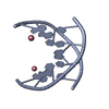
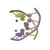
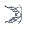
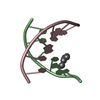
- PDB-1i7j: CRYSTAL STRUCTURE OF 2'-O-ME(CGCGCG)2: AN RNA DUPLEX AT 1.19 A RE... -
+ Open data

- Basic information
| Entry | Database: PDB / ID: 1i7j | ||||||||||||||||||
|---|---|---|---|---|---|---|---|---|---|---|---|---|---|---|---|---|---|---|---|
| Title | CRYSTAL STRUCTURE OF 2'-O-ME(CGCGCG)2: AN RNA DUPLEX AT 1.19 A RESOLUTION. 2-METHYL-2,4-PENTANEDIOL AND MAGNESIUM BINDING. | ||||||||||||||||||
 Components Components | 5'-R(* Keywords KeywordsRNA / 2'-O-MeRNA duplex / 2-methyl-2 / 4-pentanediol / magnesium / groove hydration and binding | Function / homology | RNA |  Function and homology information Function and homology informationMethod |  X-RAY DIFFRACTION / SYNCHROTRON / MOLECULAR REPLACEMENT / Resolution: 1.19 Å X-RAY DIFFRACTION / SYNCHROTRON / MOLECULAR REPLACEMENT / Resolution: 1.19 Å  Authors AuthorsAdamiak, D.A. / Rypniewski, W.R. / Milecki, J. / Adamiak, R.W. |  CitationJournal: Nucleic Acids Res. / Year: 2001 CitationJournal: Nucleic Acids Res. / Year: 2001Title: The 1.19 A X-ray structure of 2'-O-Me(CGCGCG)(2) duplex shows dehydrated RNA with 2-methyl-2,4-pentanediol in the minor groove. Authors: Adamiak, D.A. / Rypniewski, W.R. / Milecki, J. / Adamiak, R.W. #1: Journal: Nucleic Acids Res. / Year: 1997Title: CRYSTAL STRUCTURE OF 2'-O-ME(CGCGCG)2: AN RNA DUPLEX AT 1.3 A RESOLUTION. HYDRATION PATTERN OF 2'-O-METHYLATED RNA Authors: Adamiak, D.A. / Milecki, J. / Popenda, W. / Adamiak, R.W. / Dauter, A. / Rypniewski, W.R. History |
|
- Structure visualization
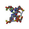
| Structure viewer | Molecule: MolmilJmol/JSmol |
|---|
- Downloads & links
-Download
| PDBx/mmCIF format | 1i7j.cif.gz | 32.1 KB | Display | PDBx/mmCIF format |
|---|---|---|---|---|
| PDB format | pdb1i7j.ent.gz | 25.3 KB | Display | PDB format |
| PDBx/mmJSON format | 1i7j.json.gz | Tree view | PDBx/mmJSON format | |
| Others |  Other downloads Other downloads |
-Validation report
| Arichive directory | https://data.pdbj.org/pub/pdb/validation_reports/i7/1i7jftp://data.pdbj.org/pub/pdb/validation_reports/i7/1i7j | HTTPS FTP |
|---|
-Related structure data
| Related structure data |  310dS S: Starting model for refinement |
|---|---|
| Similar structure data |


-Links
 PDBj
PDBj- Assembly
| Deposited unit | 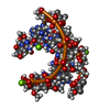
| |||||||||
|---|---|---|---|---|---|---|---|---|---|---|
| 1 |
| |||||||||
| Unit cell |
| |||||||||
| Components on special symmetry positions |
|
-Components
| #1: RNA chain | Mass: 1990.361 Da / Num. of mol.: 2 / Source method: obtained synthetically #2: Chemical | ChemComp-MG /   Mass: 24.305 Da / Num. of mol.: 5 / Source method: obtained synthetically / Formula: Mg Mass: 24.305 Da / Num. of mol.: 5 / Source method: obtained synthetically / Formula: Mg#3: Chemical |   Mass: 118.174 Da / Num. of mol.: 2 / Source method: obtained synthetically / Formula: C6H14O2 / Comment: precipitant*YM Mass: 118.174 Da / Num. of mol.: 2 / Source method: obtained synthetically / Formula: C6H14O2 / Comment: precipitant*YM#4: Water | ChemComp-HOH / |  Mass: 18.015 Da / Num. of mol.: 65 / Source method: isolated from a natural source / Formula: H2O Mass: 18.015 Da / Num. of mol.: 65 / Source method: isolated from a natural source / Formula: H2O |
|---|
-Experimental details
-Experiment
| Experiment | Method: X-RAY DIFFRACTION / Number of used crystals: 1 |
|---|
- Sample preparation
| Crystal | Density Matthews: 1.73 Å3/Da / Density % sol: 38 % | ||||||||||||||||||||||||||||||||||||
|---|---|---|---|---|---|---|---|---|---|---|---|---|---|---|---|---|---|---|---|---|---|---|---|---|---|---|---|---|---|---|---|---|---|---|---|---|---|
| Crystal grow | Temperature: 293 K / Method: vapor diffusion, hanging drop / pH: 7.5 Details: 15 mM MgCl2, 1 mM spermine tetrahydrochloride and 30-40% 2-methyl-2,4-pentanediol, pH 7.5, VAPOR DIFFUSION, HANGING DROP, temperature 293K | ||||||||||||||||||||||||||||||||||||
| Components of the solutions |
| ||||||||||||||||||||||||||||||||||||
| Crystal grow | *PLUS Temperature: 20 ℃ | ||||||||||||||||||||||||||||||||||||
| Components of the solutions | *PLUS
|
-Data collection
| Diffraction | Mean temperature: 100 K |
|---|---|
| Diffraction source | Source: SYNCHROTRON / Site: EMBL/DESY, HAMBURG  / Beamline: BW7A / Wavelength: 0.9 Å / Beamline: BW7A / Wavelength: 0.9 Å |
| Detector | Type: MARRESEARCH / Detector: IMAGE PLATE / Date: Oct 3, 1996 |
| Radiation | Monochromator: Si / Protocol: SINGLE WAVELENGTH / Monochromatic (M) / Laue (L): M / Scattering type: x-ray |
| Radiation wavelength | Wavelength: 0.9 Å / Relative weight: 1 |
| Reflection | Resolution: 1.19→20 Å / Num. obs: 8715 / % possible obs: 95.8 % / Observed criterion σ(F): 0 / Observed criterion σ(I): 0 / Redundancy: 17 % / Biso Wilson estimate: 13 Å2 / Rmerge(I) obs: 0.049 / Net I/σ(I): 23 |
| Reflection shell | Resolution: 1.19→1.21 Å / Redundancy: 2.8 % / Rmerge(I) obs: 0.46 / Mean I/σ(I) obs: 2.1 / Num. unique all: 415 / % possible all: 94 |
| Reflection | *PLUS Lowest resolution: 20 Å / Num. obs: 8715 / Num. measured all: 150605 |
| Reflection shell | *PLUS % possible obs: 94 % |
- Processing
| Software |
| ||||||||||||||||
|---|---|---|---|---|---|---|---|---|---|---|---|---|---|---|---|---|---|
| Refinement | Method to determine structure: MOLECULAR REPLACEMENT Starting model: PDB 310D Resolution: 1.19→20 Å / σ(F): 0 / σ(I): 0 / Stereochemistry target values: CCP4
| ||||||||||||||||
| Refinement step | Cycle: LAST / Resolution: 1.19→20 Å
| ||||||||||||||||
| Refine LS restraints |
| ||||||||||||||||
| LS refinement shell | Resolution: 1.19→1.21 Å /
| ||||||||||||||||
| Software | *PLUS Name: SHELXL-97 / Classification: refinement | ||||||||||||||||
| Refinement | *PLUS Lowest resolution: 20 Å / σ(F): 0 | ||||||||||||||||
| Solvent computation | *PLUS | ||||||||||||||||
| Displacement parameters | *PLUS |
