 Movie
Movie Controller

[English] 日本語
 Yorodumi
- PDB-1i78: CRYSTAL STRUCTURE OF OUTER MEMBRANE PROTEASE OMPT FROM ESCHERICHI... -
+ Open data

- Basic information
| Entry | Database: PDB / ID: 1i78 | ||||||
|---|---|---|---|---|---|---|---|
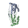
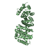
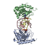
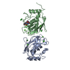
| Title | CRYSTAL STRUCTURE OF OUTER MEMBRANE PROTEASE OMPT FROM ESCHERICHIA COLI | ||||||
 Components Components | PROTEASE VII | ||||||
 Keywords Keywords | HYDROLASE / integral outer membrane protein / protease / beta barrel | ||||||
| Function / homology |  Function and homology information Function and homology informationomptin / cell outer membrane / endopeptidase activity / aspartic-type endopeptidase activity / serine-type endopeptidase activity / proteolysis Similarity search - Function | ||||||
| Biological species |  | ||||||
| Method |  X-RAY DIFFRACTION / SYNCHROTRON / SAD / Resolution: 2.6 Å X-RAY DIFFRACTION / SYNCHROTRON / SAD / Resolution: 2.6 Å | ||||||
 Authors Authors | Vandeputte-Rutten, L. / Kramer, R.A. / Kroon, J. / Dekker, N. / Egmond, M.R. / Gros, P. | ||||||
 Citation Citation | Journal: EMBO J. / Year: 2001 Title: Crystal structure of the outer membrane protease OmpT from Escherichia coli suggests a novel catalytic site. Authors: Vandeputte-Rutten, L. / Kramer, R.A. / Kroon, J. / Dekker, N. / Egmond, M.R. / Gros, P. | ||||||
| History |
|
- Structure visualization
| Structure viewer | Molecule: MolmilJmol/JSmol |
|---|
- Downloads & links
-Download
| PDBx/mmCIF format | 1i78.cif.gz | 126.3 KB | Display | PDBx/mmCIF format |
|---|---|---|---|---|
| PDB format | pdb1i78.ent.gz | 100.2 KB | Display | PDB format |
| PDBx/mmJSON format | 1i78.json.gz | Tree view | PDBx/mmJSON format | |
| Others |  Other downloads Other downloads |
-Validation report
| Arichive directory | https://data.pdbj.org/pub/pdb/validation_reports/i7/1i78ftp://data.pdbj.org/pub/pdb/validation_reports/i7/1i78 | HTTPS FTP |
|---|
-Related structure data
| Similar structure data |
|---|

-Links
 PDBj
PDBj- Assembly
| Deposited unit | 
| ||||||||
|---|---|---|---|---|---|---|---|---|---|
| 1 | 
| ||||||||
| 2 | 
| ||||||||
| 3 |
| ||||||||
| 4 | 
| ||||||||
| Unit cell |
| ||||||||
| Details | OmpT is active as a monomer. The assymetric unit contains two monomers. |
-Components
| #1: Protein | Mass: 33495.594 Da / Num. of mol.: 2 / Mutation: S99A,G216K,K217G Source method: isolated from a genetically manipulated source Source: (gene. exp.) #2: Sugar | ChemComp-BOG /   Type: D-saccharide / Mass: 292.369 Da / Num. of mol.: 4 Type: D-saccharide / Mass: 292.369 Da / Num. of mol.: 4Source method: isolated from a genetically manipulated source Formula: C14H28O6 / Comment: detergent*YM #3: Chemical |   Mass: 118.174 Da / Num. of mol.: 2 / Source method: obtained synthetically / Formula: C6H14O2 / Comment: precipitant*YM Mass: 118.174 Da / Num. of mol.: 2 / Source method: obtained synthetically / Formula: C6H14O2 / Comment: precipitant*YM#4: Water | ChemComp-HOH / |  Mass: 18.015 Da / Num. of mol.: 29 / Source method: isolated from a natural source / Formula: H2O Mass: 18.015 Da / Num. of mol.: 29 / Source method: isolated from a natural source / Formula: H2O |
|---|
-Experimental details
-Experiment
| Experiment | Method: X-RAY DIFFRACTION / Number of used crystals: 1 |
|---|
- Sample preparation
| Crystal | Density Matthews: 3.45 Å3/Da / Density % sol: 64.4 % | ||||||||||||||||||||
|---|---|---|---|---|---|---|---|---|---|---|---|---|---|---|---|---|---|---|---|---|---|
| Crystal grow | Temperature: 277 K / Method: vapor diffusion, hanging drop / pH: 5.5 Details: 30% (v/v) 2-methyl-2,4-pentanediol, 0.3 M sodium citrate PH 5.5, 1% (w/v) octyl-beta-D-glucopyranoside, VAPOR DIFFUSION, HANGING DROP, temperature 277K | ||||||||||||||||||||
| Crystal grow | *PLUS Method: unknown | ||||||||||||||||||||
| Components of the solutions | *PLUS
|
-Data collection
| Diffraction | Mean temperature: 100 K |
|---|---|
| Diffraction source | Source: SYNCHROTRON / Site: ESRF  / Beamline: ID14-4 / Wavelength: 0.9322 Å / Beamline: ID14-4 / Wavelength: 0.9322 Å |
| Detector | Type: ADSC QUANTUM 4 / Detector: CCD / Date: Dec 9, 1999 / Details: toroidal mirror |
| Radiation | Monochromator: Double crystal, Si(111) or Si(311) / Protocol: SINGLE WAVELENGTH / Monochromatic (M) / Laue (L): M / Scattering type: x-ray |
| Radiation wavelength | Wavelength: 0.9322 Å / Relative weight: 1 |
| Reflection | Resolution: 2.6→20 Å / Num. all: 29075 / Num. obs: 27400 / % possible obs: 94.3 % / Redundancy: 1.59 % / Biso Wilson estimate: 41 Å2 / Rmerge(I) obs: 0.074 / Net I/σ(I): 8.82 |
| Reflection shell | Resolution: 2.6→2.76 Å / Redundancy: 1.57 % / Rmerge(I) obs: 0.263 / Mean I/σ(I) obs: 2.54 / Num. unique all: 2766 / % possible all: 96.1 |
| Reflection | *PLUS Lowest resolution: 20 Å / Num. measured all: 43471 |
| Reflection shell | *PLUS Lowest resolution: 2.69 Å / % possible obs: 97.7 % / Num. unique obs: 2766 / Num. measured obs: 4352 |
- Processing
| Software |
| |||||||||||||||||||||||||
|---|---|---|---|---|---|---|---|---|---|---|---|---|---|---|---|---|---|---|---|---|---|---|---|---|---|---|
| Refinement | Method to determine structure: SAD / Resolution: 2.6→20 Å / Isotropic thermal model: Isotropic / Cross valid method: THROUGHOUT / Stereochemistry target values: Engh & Huber
| |||||||||||||||||||||||||
| Displacement parameters | Biso mean: 53 Å2
| |||||||||||||||||||||||||
| Refine analyze |
| |||||||||||||||||||||||||
| Refinement step | Cycle: LAST / Resolution: 2.6→20 Å
| |||||||||||||||||||||||||
| Refine LS restraints |
| |||||||||||||||||||||||||
| LS refinement shell | Resolution: 2.6→2.76 Å / Rfactor Rfree error: 0.022
| |||||||||||||||||||||||||
| Software | *PLUS Name: CNS / Classification: refinement | |||||||||||||||||||||||||
| Refinement | *PLUS Highest resolution: 2.6 Å / Lowest resolution: 20 Å / Rfactor obs: 0.238 / Rfactor Rfree: 0.28 | |||||||||||||||||||||||||
| Solvent computation | *PLUS | |||||||||||||||||||||||||
| Displacement parameters | *PLUS Biso mean: 53 Å2 | |||||||||||||||||||||||||
| Refine LS restraints | *PLUS
| |||||||||||||||||||||||||
| LS refinement shell | *PLUS Highest resolution: 2.6 Å / Rfactor Rfree: 0.36 / Rfactor Rwork: 0.318 |
