 Movie
Movie Controller


+ Open data

- Basic information
| Entry | Database: PDB / ID: 1i5b | ||||||
|---|---|---|---|---|---|---|---|
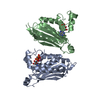
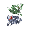
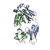
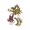
| Title | STRUCTURE OF CHEA DOMAIN P4 IN COMPLEX WITH ADPNP AND MANGANESE | ||||||
 Components Components | CHEMOTAXIS PROTEIN CHEA | ||||||
 Keywords Keywords | SIGNALING PROTEIN / TRANSFERASE / beta-alpha sandwich | ||||||
| Function / homology |  Function and homology information Function and homology informationregulation of chemotaxis / phosphorelay sensor kinase activity / histidine kinase / phosphorelay signal transduction system / chemotaxis / protein domain specific binding / ATP binding / cytoplasm Similarity search - Function | ||||||
| Biological species |   Thermotoga maritima (bacteria) Thermotoga maritima (bacteria) | ||||||
| Method |  X-RAY DIFFRACTION / SYNCHROTRON / MOLECULAR REPLACEMENT / Resolution: 1.94 Å X-RAY DIFFRACTION / SYNCHROTRON / MOLECULAR REPLACEMENT / Resolution: 1.94 Å | ||||||
 Authors Authors | Bilwes, A.M. / Quezada, C.M. / Croal, L.R. / Crane, B.R. / Simon, M.I. | ||||||
 Citation Citation | Journal: Nat.Struct.Biol. / Year: 2001 Title: Nucleotide binding by the histidine kinase CheA. Authors: Bilwes, A.M. / Quezada, C.M. / Croal, L.R. / Crane, B.R. / Simon, M.I. | ||||||
| History |
|
- Structure visualization
| Structure viewer | Molecule: MolmilJmol/JSmol |
|---|
- Downloads & links
-Download
| PDBx/mmCIF format | 1i5b.cif.gz | 88.4 KB | Display | PDBx/mmCIF format |
|---|---|---|---|---|
| PDB format | pdb1i5b.ent.gz | 66.1 KB | Display | PDB format |
| PDBx/mmJSON format | 1i5b.json.gz | Tree view | PDBx/mmJSON format | |
| Others |  Other downloads Other downloads |
-Validation report
| Arichive directory | https://data.pdbj.org/pub/pdb/validation_reports/i5/1i5bftp://data.pdbj.org/pub/pdb/validation_reports/i5/1i5b | HTTPS FTP |
|---|
-Related structure data
| Related structure data |  1i58C  1i59C  1i5aC  1i5cC  1i5dC  1b3qS S: Starting model for refinement C: citing same article ( |
|---|---|
| Similar structure data |


-Links
 PDBj
PDBj


- Assembly
| Deposited unit | 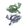
| ||||||||||
|---|---|---|---|---|---|---|---|---|---|---|---|
| 1 |
| ||||||||||
| Unit cell |
|
-Components
| #1: Protein | Mass: 21079.270 Da / Num. of mol.: 2 / Fragment: DOMAIN P4 / Mutation: R354H, I353S, K352G Source method: isolated from a genetically manipulated source Source: (gene. exp.) Thermotoga maritima (bacteria) / Species (production host): Escherichia coli / Production host: References: UniProt: Q56310, Transferases; Transferring phosphorus-containing groups; Phosphotransferases with a nitrogenous group as acceptor #2: Chemical | ChemComp-ACT / |   Mass: 59.044 Da / Num. of mol.: 1 / Source method: obtained synthetically / Formula: C2H3O2 Mass: 59.044 Da / Num. of mol.: 1 / Source method: obtained synthetically / Formula: C2H3O2#3: Chemical |   Mass: 54.938 Da / Num. of mol.: 2 / Source method: obtained synthetically / Formula: Mn Mass: 54.938 Da / Num. of mol.: 2 / Source method: obtained synthetically / Formula: Mn#4: Chemical |   Mass: 506.196 Da / Num. of mol.: 2 / Source method: obtained synthetically / Formula: C10H17N6O12P3 / Comment: AMP-PNP, energy-carrying molecule analogue*YM Mass: 506.196 Da / Num. of mol.: 2 / Source method: obtained synthetically / Formula: C10H17N6O12P3 / Comment: AMP-PNP, energy-carrying molecule analogue*YM#5: Water | ChemComp-HOH / |  Mass: 18.015 Da / Num. of mol.: 193 / Source method: isolated from a natural source / Formula: H2O Mass: 18.015 Da / Num. of mol.: 193 / Source method: isolated from a natural source / Formula: H2O |
|---|
-Experimental details
-Experiment
| Experiment | Method: X-RAY DIFFRACTION / Number of used crystals: 1 |
|---|
- Sample preparation
| Crystal | Density Matthews: 2.02 Å3/Da / Density % sol: 39.01 % | ||||||||||||||||||||||||||||||||||||||||||
|---|---|---|---|---|---|---|---|---|---|---|---|---|---|---|---|---|---|---|---|---|---|---|---|---|---|---|---|---|---|---|---|---|---|---|---|---|---|---|---|---|---|---|---|
| Crystal grow | Temperature: 293 K / Method: vapor diffusion, hanging drop / pH: 5 Details: PEG 8000 33-36% Ammonium acetate 0.8 M sodium acetate 0.085 M pH 4.5. VAPOR DIFFUSION, HANGING DROP at 293 K | ||||||||||||||||||||||||||||||||||||||||||
| Crystal grow | *PLUS Temperature: 25 ℃ / pH: 4.5 / Method: vapor diffusion | ||||||||||||||||||||||||||||||||||||||||||
| Components of the solutions | *PLUS
|
-Data collection
| Diffraction | Mean temperature: 100 K |
|---|---|
| Diffraction source | Source: SYNCHROTRON / Site: SSRL  / Beamline: BL9-2 / Wavelength: 1.89 Å / Beamline: BL9-2 / Wavelength: 1.89 Å |
| Detector | Type: ADSC QUANTUM 4 / Detector: CCD / Date: May 10, 2000 |
| Radiation | Protocol: SINGLE WAVELENGTH / Monochromatic (M) / Laue (L): M / Scattering type: x-ray |
| Radiation wavelength | Wavelength: 1.89 Å / Relative weight: 1 |
| Reflection | Resolution: 1.94→30 Å / Num. all: 36885 / Num. obs: 36885 / % possible obs: 80 % / Observed criterion σ(I): 1 / Redundancy: 2 % / Biso Wilson estimate: 24.9 Å2 / Rmerge(I) obs: 0.057 / Net I/σ(I): 30 |
| Reflection shell | Resolution: 1.94→2.02 Å / Redundancy: 2 % / Rmerge(I) obs: 0.143 / Mean I/σ(I) obs: 10 / % possible all: 15 |
| Reflection | *PLUS % possible obs: 80 % |
| Reflection shell | *PLUS % possible obs: 15 % |
- Processing
| Software |
| ||||||||||||||||||||||||||||||||||||||||
|---|---|---|---|---|---|---|---|---|---|---|---|---|---|---|---|---|---|---|---|---|---|---|---|---|---|---|---|---|---|---|---|---|---|---|---|---|---|---|---|---|---|
| Refinement | Method to determine structure: MOLECULAR REPLACEMENT Starting model: 1B3Q Resolution: 1.94→19.75 Å / Rfactor Rfree error: 0.005 / Data cutoff high absF: 79648775.42 / Data cutoff low absF: 0 / Isotropic thermal model: RESTRAINED / Cross valid method: THROUGHOUT / σ(F): 0 / σ(I): 0 / Stereochemistry target values: Engh & Huber
| ||||||||||||||||||||||||||||||||||||||||
| Solvent computation | Solvent model: FLAT MODEL / Bsol: 42.55 Å2 / ksol: 0.35 e/Å3 | ||||||||||||||||||||||||||||||||||||||||
| Displacement parameters | Biso mean: 25.6 Å2
| ||||||||||||||||||||||||||||||||||||||||
| Refine analyze |
| ||||||||||||||||||||||||||||||||||||||||
| Refinement step | Cycle: LAST / Resolution: 1.94→19.75 Å
| ||||||||||||||||||||||||||||||||||||||||
| Refine LS restraints |
| ||||||||||||||||||||||||||||||||||||||||
| LS refinement shell | Resolution: 1.95→2.02 Å / Total num. of bins used: 10 /
| ||||||||||||||||||||||||||||||||||||||||
| Xplor file |
| ||||||||||||||||||||||||||||||||||||||||
| Software | *PLUS Name: CNS / Version: 1 / Classification: refinement | ||||||||||||||||||||||||||||||||||||||||
| Refinement | *PLUS σ(F): 0 / % reflection Rfree: 9.5 % | ||||||||||||||||||||||||||||||||||||||||
| Solvent computation | *PLUS | ||||||||||||||||||||||||||||||||||||||||
| Displacement parameters | *PLUS Biso mean: 25.6 Å2 | ||||||||||||||||||||||||||||||||||||||||
| Refine LS restraints | *PLUS
| ||||||||||||||||||||||||||||||||||||||||
| LS refinement shell | *PLUS Rfactor Rfree: 0.25 / Rfactor Rwork: 0.27 |
