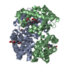
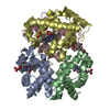
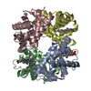
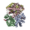
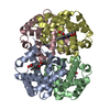
 Movie
Movie Controller


+ Open data

- Basic information
| Entry | Database: PDB / ID: 1i3d | ||||||
|---|---|---|---|---|---|---|---|
| Title | HUMAN CARBONMONOXY HEMOGLOBIN BART'S (GAMMA4) | ||||||
 Components Components | HEMOGLOBIN GAMMA CHAINS | ||||||
 Keywords Keywords | OXYGEN STORAGE/TRANSPORT / OXYGEN TRANSPORT / OXYGEN STORAGE-TRANSPORT COMPLEX | ||||||
| Function / homology |  Function and homology information Function and homology informationhemoglobin alpha binding / haptoglobin-hemoglobin complex / hemoglobin complex / oxygen transport / erythrocyte development / oxygen carrier activity / carbon dioxide transport / oxygen binding / Factors involved in megakaryocyte development and platelet production / heme binding ...hemoglobin alpha binding / haptoglobin-hemoglobin complex / hemoglobin complex / oxygen transport / erythrocyte development / oxygen carrier activity / carbon dioxide transport / oxygen binding / Factors involved in megakaryocyte development and platelet production / heme binding / metal ion binding / cytosol Similarity search - Function | ||||||
| Biological species |  Homo sapiens (human) Homo sapiens (human) | ||||||
| Method |  X-RAY DIFFRACTION / SYNCHROTRON / MOLECULAR REPLACEMENT / Resolution: 1.7 Å X-RAY DIFFRACTION / SYNCHROTRON / MOLECULAR REPLACEMENT / Resolution: 1.7 Å | ||||||
 Authors Authors | Kidd, R.D. / Baker, H.M. / Mathews, A.J. / Brittain, T. / Baker, E.N. | ||||||
 Citation Citation | Journal: Protein Sci. / Year: 2001 Title: Oligomerization and ligand binding in a homotetrameric hemoglobin: two high-resolution crystal structures of hemoglobin Bart's (gamma(4)), a marker for alpha-thalassemia. Authors: Kidd, R.D. / Baker, H.M. / Mathews, A.J. / Brittain, T. / Baker, E.N. | ||||||
| History |
|
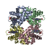
- Structure visualization
| Structure viewer | Molecule: MolmilJmol/JSmol |
|---|
- Downloads & links
-Download
| PDBx/mmCIF format | 1i3d.cif.gz | 81.8 KB | Display | PDBx/mmCIF format |
|---|---|---|---|---|
| PDB format | pdb1i3d.ent.gz | 59.9 KB | Display | PDB format |
| PDBx/mmJSON format | 1i3d.json.gz | Tree view | PDBx/mmJSON format | |
| Others |  Other downloads Other downloads |
-Validation report
| Arichive directory | https://data.pdbj.org/pub/pdb/validation_reports/i3/1i3dftp://data.pdbj.org/pub/pdb/validation_reports/i3/1i3d | HTTPS FTP |
|---|
-Related structure data
| Related structure data | 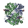 1i3eC 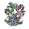 1cbmS C: citing same article ( S: Starting model for refinement |
|---|---|
| Similar structure data |
-Links
 PDBj
PDBj


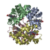
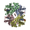
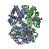
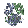
- Assembly
| Deposited unit | 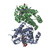
| ||||||||
|---|---|---|---|---|---|---|---|---|---|
| 1 | 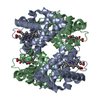
| ||||||||
| Unit cell |
| ||||||||
| Details | The second globin dimer of the homotetramer is generated by the two fold axis: -x, -y+1, z. |
-Components
| #1: Protein | Mass: 16031.255 Da / Num. of mol.: 2 Source method: isolated from a genetically manipulated source Source: (gene. exp.) Homo sapiens (human) / Gene: HBG1 / Plasmid: PRMAE389 / Production host:  #2: Chemical |   Mass: 616.487 Da / Num. of mol.: 2 / Source method: obtained synthetically / Formula: C34H32FeN4O4 Mass: 616.487 Da / Num. of mol.: 2 / Source method: obtained synthetically / Formula: C34H32FeN4O4#3: Chemical |   Mass: 28.010 Da / Num. of mol.: 2 / Source method: obtained synthetically / Formula: CO Mass: 28.010 Da / Num. of mol.: 2 / Source method: obtained synthetically / Formula: CO#4: Water | ChemComp-HOH / |  Mass: 18.015 Da / Num. of mol.: 286 / Source method: isolated from a natural source / Formula: H2O Mass: 18.015 Da / Num. of mol.: 286 / Source method: isolated from a natural source / Formula: H2O |
|---|
-Experimental details
-Experiment
| Experiment | Method: X-RAY DIFFRACTION / Number of used crystals: 1 |
|---|
- Sample preparation
| Crystal | Density Matthews: 2.1 Å3/Da / Density % sol: 41.4 % | ||||||||||||||||||||||||||||||
|---|---|---|---|---|---|---|---|---|---|---|---|---|---|---|---|---|---|---|---|---|---|---|---|---|---|---|---|---|---|---|---|
| Crystal grow | Temperature: 291 K / Method: vapor diffusion, hanging drop / pH: 4.9 Details: acetic acid/KOH, MePEG 5000, pH 4.9, VAPOR DIFFUSION, HANGING DROP, temperature 291K | ||||||||||||||||||||||||||||||
| Crystal grow | *PLUS Temperature: 18 ℃ / pH: 7 | ||||||||||||||||||||||||||||||
| Components of the solutions | *PLUS
|
-Data collection
| Diffraction | Mean temperature: 110 K |
|---|---|
| Diffraction source | Source: SYNCHROTRON / Site: SSRL  / Beamline: BL7-1 / Wavelength: 1.08 Å / Beamline: BL7-1 / Wavelength: 1.08 Å |
| Detector | Type: MAR scanner 345 mm plate / Detector: IMAGE PLATE / Date: Dec 10, 1998 / Details: mirror |
| Radiation | Monochromator: Cyclindrically bent triangular Si(111) asymmetric cut, horizontal focus Protocol: SINGLE WAVELENGTH / Monochromatic (M) / Laue (L): M / Scattering type: x-ray |
| Radiation wavelength | Wavelength: 1.08 Å / Relative weight: 1 |
| Reflection | Resolution: 1.7→30 Å / Num. all: 27848 / Num. obs: 27848 / % possible obs: 91.5 % / Observed criterion σ(F): 0 / Redundancy: 3.8 % / Biso Wilson estimate: 17.4 Å2 / Rmerge(I) obs: 0.078 / Net I/σ(I): 8.7 |
| Reflection shell | Resolution: 1.7→1.76 Å / Redundancy: 3.7 % / Rmerge(I) obs: 0.283 / Mean I/σ(I) obs: 3 / Num. unique all: 2942 / % possible all: 98.5 |
| Reflection | *PLUS Num. measured all: 105161 |
| Reflection shell | *PLUS % possible obs: 98.5 % |
- Processing
| Software |
| |||||||||||||||||||||||||
|---|---|---|---|---|---|---|---|---|---|---|---|---|---|---|---|---|---|---|---|---|---|---|---|---|---|---|
| Refinement | Method to determine structure: MOLECULAR REPLACEMENT Starting model: PDB ENTRY 1CBM Resolution: 1.7→20 Å / Rfactor Rfree error: 0.006 / Isotropic thermal model: Overall / Cross valid method: THROUGHOUT / σ(F): 0 / Stereochemistry target values: Engh & Huber
| |||||||||||||||||||||||||
| Solvent computation | Solvent model: FLAT MODEL / Bsol: 32.1634 Å2 / ksol: 0.310647 e/Å3 | |||||||||||||||||||||||||
| Displacement parameters | Biso mean: 16.9 Å2
| |||||||||||||||||||||||||
| Refine analyze |
| |||||||||||||||||||||||||
| Refinement step | Cycle: LAST / Resolution: 1.7→20 Å
| |||||||||||||||||||||||||
| Refine LS restraints |
| |||||||||||||||||||||||||
| LS refinement shell | Resolution: 1.7→1.81 Å / Rfactor Rfree error: 0.018 / Total num. of bins used: 6
| |||||||||||||||||||||||||
| Xplor file |
| |||||||||||||||||||||||||
| Software | *PLUS Name: CNS / Version: 1 / Classification: refinement | |||||||||||||||||||||||||
| Refinement | *PLUS Lowest resolution: 20 Å / σ(F): 0 / % reflection Rfree: 10 % / Rfactor obs: 0.211 | |||||||||||||||||||||||||
| Solvent computation | *PLUS | |||||||||||||||||||||||||
| Displacement parameters | *PLUS Biso mean: 16.9 Å2 | |||||||||||||||||||||||||
| Refine LS restraints | *PLUS
| |||||||||||||||||||||||||
| LS refinement shell | *PLUS Rfactor Rfree: 0.376 / % reflection Rfree: 9.8 % / Rfactor Rwork: 0.332 |
