 Movie
Movie Controller

[English] 日本語
 Yorodumi
- PDB-1i2d: CRYSTAL STRUCTURE OF ATP SULFURYLASE FROM PENICILLIUM CHRYSOGENUM -
+ Open data

- Basic information
| Entry | Database: PDB / ID: 1i2d | ||||||
|---|---|---|---|---|---|---|---|
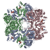
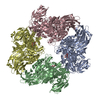
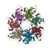
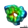
| Title | CRYSTAL STRUCTURE OF ATP SULFURYLASE FROM PENICILLIUM CHRYSOGENUM | ||||||
 Components Components | ATP SULFURYLASE | ||||||
 Keywords Keywords | TRANSFERASE / nucleotide binding / allosteric / hexamer | ||||||
| Function / homology |  Function and homology information Function and homology informationsulfate assimilation via adenylyl sulfate reduction / sulfate assimilation, phosphoadenylyl sulfate reduction by phosphoadenylyl-sulfate reductase (thioredoxin) / sulfate adenylyltransferase / adenylylsulfate kinase activity / sulfate adenylyltransferase (ATP) activity / hydrogen sulfide biosynthetic process / ATP binding / cytoplasm Similarity search - Function | ||||||
| Biological species |  Penicillium chrysogenum (fungus) Penicillium chrysogenum (fungus) | ||||||
| Method |  X-RAY DIFFRACTION / SYNCHROTRON / MAD / Resolution: 2.81 Å X-RAY DIFFRACTION / SYNCHROTRON / MAD / Resolution: 2.81 Å | ||||||
 Authors Authors | MacRae, I.J. / Segel, I.H. / Fisher, A.J. | ||||||
 Citation Citation | Journal: Biochemistry / Year: 2001 Title: Crystal structure of ATP sulfurylase from Penicillium chrysogenum: insights into the allosteric regulation of sulfate assimilation. Authors: MacRae, I.J. / Segel, I.H. / Fisher, A.J. | ||||||
| History |
|
- Structure visualization
| Structure viewer | Molecule: MolmilJmol/JSmol |
|---|
- Downloads & links
-Download
| PDBx/mmCIF format | 1i2d.cif.gz | 347.5 KB | Display | PDBx/mmCIF format |
|---|---|---|---|---|
| PDB format | pdb1i2d.ent.gz | 282.8 KB | Display | PDB format |
| PDBx/mmJSON format | 1i2d.json.gz | Tree view | PDBx/mmJSON format | |
| Others |  Other downloads Other downloads |
-Validation report
| Summary document | 1i2d_validation.pdf.gz | 719.3 KB | Display | wwPDB validaton report |
|---|---|---|---|---|
| Full document | 1i2d_full_validation.pdf.gz | 772.5 KB | Display | |
| Data in XML | 1i2d_validation.xml.gz | 41 KB | Display | |
| Data in CIF | 1i2d_validation.cif.gz | 62.2 KB | Display | |
| Arichive directory | https://data.pdbj.org/pub/pdb/validation_reports/i2/1i2dftp://data.pdbj.org/pub/pdb/validation_reports/i2/1i2d | HTTPS FTP |
-Related structure data
| Similar structure data |
|---|


-Links
 PDBj
PDBj
- Assembly
| Deposited unit | 
| ||||||||
|---|---|---|---|---|---|---|---|---|---|
| 1 | 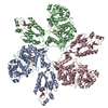
| ||||||||
| Unit cell |
| ||||||||
| Details | The biological assembly is a hexamer generated from the trimer in the asymmetric unit by the operation: -x, -y+1, z. |
-Components
| #1: Protein | Mass: 64048.469 Da / Num. of mol.: 3 Source method: isolated from a genetically manipulated source Source: (gene. exp.) Penicillium chrysogenum (fungus) / Gene: APS / Plasmid: PET23A / Species (production host): Escherichia coli / Production host:  #2: Chemical | ChemComp-ADX /   Type: RNA linking / Mass: 427.284 Da / Num. of mol.: 6 / Source method: obtained synthetically / Formula: C10H14N5O10PS Type: RNA linking / Mass: 427.284 Da / Num. of mol.: 6 / Source method: obtained synthetically / Formula: C10H14N5O10PS#3: Water | ChemComp-HOH / |  Mass: 18.015 Da / Num. of mol.: 282 / Source method: isolated from a natural source / Formula: H2O Mass: 18.015 Da / Num. of mol.: 282 / Source method: isolated from a natural source / Formula: H2ONonpolymer details | ADX 574 AND 575 ARE ASSOCIATED WITH CHAIN A. ADX 576 AND 577 ARE ASSOCIATED WITH CHAIN B. ADX 578 ...ADX 574 AND 575 ARE ASSOCIATED | Sequence details | THE SEQUENCE/SEQUENCE DATABASE CONFLICT IS DUE TO AN ERROR IN THE DATABASE SEQUENCE. THE AUTHORS ...THE SEQUENCE/SEQUENCE DATABASE CONFLICT IS DUE TO AN ERROR IN THE DATABASE SEQUENCE. THE AUTHORS HAVE SEQUENCED THE GENE 4 TIMES AND DETERMINED | |
|---|
-Experimental details
-Experiment
| Experiment | Method: X-RAY DIFFRACTION / Number of used crystals: 1 |
|---|
- Sample preparation
| Crystal | Density Matthews: 3.9 Å3/Da / Density % sol: 68.49 % | |||||||||||||||||||||||||
|---|---|---|---|---|---|---|---|---|---|---|---|---|---|---|---|---|---|---|---|---|---|---|---|---|---|---|
| Crystal grow | Temperature: 277 K / Method: vapor diffusion, hanging drop / pH: 8 Details: 1 M sodium citrate, 2% PEG 400, 100 mM Tris-Cl, 5 mM adenosine 5'-phosphosulfate, pH 8.0, VAPOR DIFFUSION, HANGING DROP, temperature 277K | |||||||||||||||||||||||||
| Crystal | *PLUS Density % sol: 69 % | |||||||||||||||||||||||||
| Crystal grow | *PLUS Temperature: 4 ℃ | |||||||||||||||||||||||||
| Components of the solutions | *PLUS
|
-Data collection
| Diffraction | Mean temperature: 100 K |
|---|---|
| Diffraction source | Source: SYNCHROTRON / Site: SSRL  / Beamline: BL7-1 / Wavelength: 1.08 Å / Beamline: BL7-1 / Wavelength: 1.08 Å |
| Detector | Type: MARRESEARCH / Detector: IMAGE PLATE / Date: Apr 15, 2000 |
| Radiation | Monochromator: synchrotron / Protocol: SINGLE WAVELENGTH / Monochromatic (M) / Laue (L): M / Scattering type: x-ray |
| Radiation wavelength | Wavelength: 1.08 Å / Relative weight: 1 |
| Reflection | Resolution: 2.8→30 Å / Num. all: 75367 / Num. obs: 75367 / % possible obs: 95.9 % / Observed criterion σ(F): -3 / Observed criterion σ(I): -3 / Redundancy: 3.4 % / Biso Wilson estimate: 60.4 Å2 / Rmerge(I) obs: 0.064 / Net I/σ(I): 14 |
| Reflection shell | Resolution: 2.8→2.9 Å / Redundancy: 2.8 % / Rmerge(I) obs: 0.354 / Mean I/σ(I) obs: 2.8 / Num. unique all: 5681 / % possible all: 73 |
| Reflection | *PLUS Num. measured all: 257680 |
- Processing
| Software |
| ||||||||||||||||||||||||||||||||||||||||
|---|---|---|---|---|---|---|---|---|---|---|---|---|---|---|---|---|---|---|---|---|---|---|---|---|---|---|---|---|---|---|---|---|---|---|---|---|---|---|---|---|---|
| Refinement | Method to determine structure: MAD / Resolution: 2.81→29.08 Å / Rfactor Rfree error: 0.004 / Data cutoff high absF: 2928114.36 / Data cutoff low absF: 0 / Isotropic thermal model: RESTRAINED / Cross valid method: THROUGHOUT / σ(F): 0 / σ(I): 0 / Stereochemistry target values: Engh & Huber Details: Non-crystallographic symmetry was tightly imposed. CYS 42 is the only CYS that appears to have HG bound.
| ||||||||||||||||||||||||||||||||||||||||
| Solvent computation | Solvent model: FLAT MODEL / Bsol: 50.04 Å2 / ksol: 0.318 e/Å3 | ||||||||||||||||||||||||||||||||||||||||
| Displacement parameters | Biso mean: 65.6 Å2
| ||||||||||||||||||||||||||||||||||||||||
| Refine analyze |
| ||||||||||||||||||||||||||||||||||||||||
| Refinement step | Cycle: LAST / Resolution: 2.81→29.08 Å
| ||||||||||||||||||||||||||||||||||||||||
| Refine LS restraints |
| ||||||||||||||||||||||||||||||||||||||||
| LS refinement shell | Resolution: 2.8→2.98 Å / Rfactor Rfree error: 0.017 / Total num. of bins used: 6
| ||||||||||||||||||||||||||||||||||||||||
| Xplor file |
| ||||||||||||||||||||||||||||||||||||||||
| Software | *PLUS Name: CNS / Version: 1 / Classification: refinement | ||||||||||||||||||||||||||||||||||||||||
| Refinement | *PLUS σ(F): 0 / % reflection Rfree: 5.1 % | ||||||||||||||||||||||||||||||||||||||||
| Solvent computation | *PLUS | ||||||||||||||||||||||||||||||||||||||||
| Displacement parameters | *PLUS Biso mean: 65.6 Å2 | ||||||||||||||||||||||||||||||||||||||||
| Refine LS restraints | *PLUS
| ||||||||||||||||||||||||||||||||||||||||
| LS refinement shell | *PLUS Rfactor Rfree: 0.382 / % reflection Rfree: 4.9 % / Rfactor Rwork: 0.323 |
