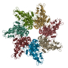
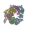
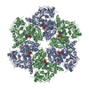
- PDB-6uji: Low resolution crystal structure (5.5 A) of the anthrax toxin pro... -
+
Open data
ID or keywords:
Loading...
-
Basic information
Entry
Database: PDB / ID: 6uji
Title
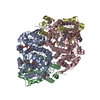
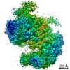
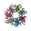
Low resolution crystal structure (5.5 A) of the anthrax toxin protective antigen heptamer prepore D425A mutant
Components
Protective antigen PA-63
Keywords
TOXIN / Anthrax Toxin / PA63 heptamer
Function / homology
Function and homology information
symbiont-mediated suppression of host MAPK cascade / host cell cytosol / Uptake and function of anthrax toxins / host cell endosome membrane / protein homooligomerization / toxin activity / host cell plasma membrane / extracellular region / metal ion binding / identical protein binding Similarity search - Function
National Institutes of Health/National Institute of General Medical Sciences (NIH/NIGMS)
P30 GM110761
United States
Citation
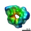
Journal: J Mol Biol / Year: 2022 Title: Structure of the Anthrax Protective Antigen D425A Dominant Negative Mutant Reveals a Stalled Intermediate State of Pore Maturation. Authors: Harry Scott / Wei Huang / Kiran Andra / Sireesha Mamillapalli / Srinivas Gonti / Alexander Day / Kaiming Zhang / Nurjahan Mehzabeen / Kevin P Battaile / Anjali Raju / Scott Lovell / James G ...Authors: Harry Scott / Wei Huang / Kiran Andra / Sireesha Mamillapalli / Srinivas Gonti / Alexander Day / Kaiming Zhang / Nurjahan Mehzabeen / Kevin P Battaile / Anjali Raju / Scott Lovell / James G Bann / Derek J Taylor / Abstract: The tripartite protein complex produced by anthrax bacteria (Bacillus anthracis) is a member of the AB family of β-barrel pore-forming toxins. The protective antigen (PA) component forms an ...The tripartite protein complex produced by anthrax bacteria (Bacillus anthracis) is a member of the AB family of β-barrel pore-forming toxins. The protective antigen (PA) component forms an oligomeric prepore that assembles on the host cell surface and serves as a scaffold for binding of lethal and edema factors. Following endocytosis, the acidic environment of the late endosome triggers a pH-induced conformational rearrangement to promote maturation of the PA prepore to a functional, membrane spanning pore that facilitates delivery of lethal and edema factors to the cytosol of the infected host. Here, we show that the dominant-negative D425A mutant of PA stalls anthrax pore maturation in an intermediate state at acidic pH. Our 2.7 Å cryo-EM structure of the intermediate state reveals structural rearrangements that involve constriction of the oligomeric pore combined with an intramolecular dissociation of the pore-forming module. In addition to defining the early stages of anthrax pore maturation, the structure identifies asymmetric conformational changes in the oligomeric pore that are influenced by the precise configuration of adjacent protomers.
Resolution: 5.5→44.83 Å / Cor.coef. Fo:Fc: 0.898 / Cor.coef. Fo:Fc free: 0.873 / Cross valid method: THROUGHOUT / σ(F): 0 / SU Rfree Blow DPI: 1.587 Details: Rigid body refinement with B-factors set to the Wilson B-factor
In the structure databanks used in Yorodumi, some data are registered as the other names, "COVID-19 virus" and "2019-nCoV". Here are the details of the virus and the list of structure data.
Jan 31, 2019. EMDB accession codes are about to change! (news from PDBe EMDB page)
EMDB accession codes are about to change! (news from PDBe EMDB page)
The allocation of 4 digits for EMDB accession codes will soon come to an end. Whilst these codes will remain in use, new EMDB accession codes will include an additional digit and will expand incrementally as the available range of codes is exhausted. The current 4-digit format prefixed with “EMD-” (i.e. EMD-XXXX) will advance to a 5-digit format (i.e. EMD-XXXXX), and so on. It is currently estimated that the 4-digit codes will be depleted around Spring 2019, at which point the 5-digit format will come into force.
The EM Navigator/Yorodumi systems omit the EMD- prefix.
Related info.:Q: What is EMD? / ID/Accession-code notation in Yorodumi/EM Navigator
Yorodumi is a browser for structure data from EMDB, PDB, SASBDB, etc.
This page is also the successor to EM Navigator detail page, and also detail information page/front-end page for Omokage search.
The word "yorodu" (or yorozu) is an old Japanese word meaning "ten thousand". "mi" (miru) is to see.
Related info.:EMDB / PDB / SASBDB / Comparison of 3 databanks / Yorodumi Search / Aug 31, 2016. New EM Navigator & Yorodumi / Yorodumi Papers / Jmol/JSmol / Function and homology information / Changes in new EM Navigator and Yorodumi
 Movie
Movie Controller

 Yorodumi
 Open data
 Basic information
 Components
 Keywords
 Function and homology information
 X-RAY DIFFRACTION /
  Authors
 United States, 1items
  Citation
 Structure visualization
 Downloads & links
 Other downloads


 PDBj
PDBj


 Assembly
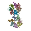

 Sample preparation
 Processing