 Movie
Movie Controller

[English] 日本語
 Yorodumi
- PDB-1i1a: CRYSTAL STRUCTURE OF THE NEONATAL FC RECEPTOR COMPLEXED WITH A HE... -
+ Open data

- Basic information
| Entry | Database: PDB / ID: 1i1a | |||||||||
|---|---|---|---|---|---|---|---|---|---|---|
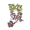
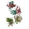
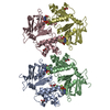
| Title | CRYSTAL STRUCTURE OF THE NEONATAL FC RECEPTOR COMPLEXED WITH A HETERODIMERIC FC | |||||||||
 Components Components |
| |||||||||
 Keywords Keywords | IMMUNE SYSTEM / MHC class I fold / Ig constant domains | |||||||||
| Function / homology |  Function and homology information Function and homology informationEndosomal/Vacuolar pathway / DAP12 interactions / Immunoregulatory interactions between a Lymphoid and a non-Lymphoid cell / Antigen Presentation: Folding, assembly and peptide loading of class I MHC / DAP12 signaling / ER-Phagosome pathway / IgG receptor activity / IgG binding / immunoglobulin complex, circulating / Neutrophil degranulation ...Endosomal/Vacuolar pathway / DAP12 interactions / Immunoregulatory interactions between a Lymphoid and a non-Lymphoid cell / Antigen Presentation: Folding, assembly and peptide loading of class I MHC / DAP12 signaling / ER-Phagosome pathway / IgG receptor activity / IgG binding / immunoglobulin complex, circulating / Neutrophil degranulation / immunoglobulin receptor binding / regulation of membrane depolarization / humoral immune response / beta-2-microglobulin binding / complement activation, classical pathway / response to cadmium ion / antigen binding / regulation of iron ion transport / cellular response to iron(III) ion / negative regulation of iron ion transport / negative regulation of forebrain neuron differentiation / antigen processing and presentation of exogenous protein antigen via MHC class Ib, TAP-dependent / iron ion transport / peptide antigen assembly with MHC class I protein complex / regulation of erythrocyte differentiation / response to molecule of bacterial origin / HFE-transferrin receptor complex / MHC class I peptide loading complex / transferrin transport / cellular response to iron ion / negative regulation of receptor-mediated endocytosis / positive regulation of T cell cytokine production / antigen processing and presentation of endogenous peptide antigen via MHC class I / MHC class I protein complex / peptide antigen assembly with MHC class II protein complex / negative regulation of neurogenesis / cellular response to nicotine / MHC class II protein complex / positive regulation of receptor-mediated endocytosis / multicellular organismal-level iron ion homeostasis / positive regulation of T cell mediated cytotoxicity / antigen processing and presentation of exogenous peptide antigen via MHC class II / positive regulation of immune response / peptide antigen binding / positive regulation of T cell activation / negative regulation of epithelial cell proliferation / sensory perception of smell / positive regulation of cellular senescence / MHC class II protein complex binding / T cell differentiation in thymus / late endosome membrane / antimicrobial humoral immune response mediated by antimicrobial peptide / negative regulation of neuron projection development / antibacterial humoral response / protein refolding / cellular response to lipopolysaccharide / amyloid fibril formation / protein homotetramerization / intracellular iron ion homeostasis / learning or memory / endosome membrane / immune response / response to xenobiotic stimulus / external side of plasma membrane / innate immune response / lysosomal membrane / structural molecule activity / Golgi apparatus / protein homodimerization activity / : / identical protein binding / plasma membrane / cytosol Similarity search - Function | |||||||||
| Biological species |  | |||||||||
| Method |  X-RAY DIFFRACTION / SYNCHROTRON / MOLECULAR REPLACEMENT / Resolution: 2.8 Å X-RAY DIFFRACTION / SYNCHROTRON / MOLECULAR REPLACEMENT / Resolution: 2.8 Å | |||||||||
 Authors Authors | Martin, W.L. / West Jr., A.P. / Gan, L. / Bjorkman, P.J. | |||||||||
 Citation Citation | Journal: Mol.Cell / Year: 2001 Title: Crystal structure at 2.8 A of an FcRn/heterodimeric Fc complex: mechanism of pH-dependent binding. Authors: Martin, W.L. / West Jr., A.P. / Gan, L. / Bjorkman, P.J. | |||||||||
| History |
|
- Structure visualization
| Structure viewer | Molecule: MolmilJmol/JSmol |
|---|
- Downloads & links
-Download
| PDBx/mmCIF format | 1i1a.cif.gz | 180 KB | Display | PDBx/mmCIF format |
|---|---|---|---|---|
| PDB format | pdb1i1a.ent.gz | 140.6 KB | Display | PDB format |
| PDBx/mmJSON format | 1i1a.json.gz | Tree view | PDBx/mmJSON format | |
| Others |  Other downloads Other downloads |
-Validation report
| Arichive directory | https://data.pdbj.org/pub/pdb/validation_reports/i1/1i1aftp://data.pdbj.org/pub/pdb/validation_reports/i1/1i1a | HTTPS FTP |
|---|
-Related structure data
| Related structure data | 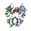 1i1cC  1fc1S  3fruS S: Starting model for refinement C: citing same article ( |
|---|---|
| Similar structure data |


-Links
 PDBj
PDBj


- Assembly
| Deposited unit | 
| ||||||||
|---|---|---|---|---|---|---|---|---|---|
| 1 |
| ||||||||
| Unit cell |
| ||||||||
| Details | The glycosylated heterodimer is the biological unit. The cysteines attached to cysteines 48 and 226 are the result of long term storage in the growth media. / The biological unit is a glycosylated homodimer. One of the two amino acid chains has been mutated as noted making the biological unit in this structure a heterodimer |
-Components
-Protein , 2 types, 2 molecules AB
| #1: Protein | Mass: 30322.887 Da / Num. of mol.: 1 / Fragment: EXTRACELLULAR DOMAIN Source method: isolated from a genetically manipulated source Source: (gene. exp.)  Cricetulus griseus (Chinese hamster) / Strain (production host): CHO-K1 / References: UniProt: P13599 Cricetulus griseus (Chinese hamster) / Strain (production host): CHO-K1 / References: UniProt: P13599 |
|---|---|
| #2: Protein | Mass: 11652.282 Da / Num. of mol.: 1 / Fragment: FC FRAGMENT Source method: isolated from a genetically manipulated source Source: (gene. exp.) Cricetulus griseus (Chinese hamster) / Strain (production host): CHO-K1 / References: UniProt: P07151 |
-IG GAMMA-2A CHAIN C ... , 2 types, 2 molecules CD
| #3: Protein | Mass: 25248.506 Da / Num. of mol.: 1 / Fragment: WILD-TYPE FC FRAGMENT Source method: isolated from a genetically manipulated source Source: (gene. exp.) Cricetulus griseus (Chinese hamster) / Strain (production host): CHO-K1 / References: UniProt: P20760 |
|---|---|
| #4: Protein | Mass: 26650.855 Da / Num. of mol.: 1 / Fragment: NON-FCRN-BINDING FC FRAGMENT / Mutation: T252G, I253G, T254G, H310E, H433E, H435E Source method: isolated from a genetically manipulated source Source: (gene. exp.) Cricetulus griseus (Chinese hamster) / Strain (production host): CHO-K1 / References: UniProt: P20760 |
-Sugars , 5 types, 6 molecules 
| #5: Polysaccharide | alpha-L-fucopyranose-(1-6)-2-acetamido-2-deoxy-beta-D-glucopyranose Source method: isolated from a genetically manipulated source |
|---|---|
| #6: Polysaccharide | 2-acetamido-2-deoxy-alpha-D-glucopyranose-(1-2)-alpha-D-mannopyranose-(1-3)-beta-D-mannopyranose-(1- ...2-acetamido-2-deoxy-alpha-D-glucopyranose-(1-2)-alpha-D-mannopyranose-(1-3)-beta-D-mannopyranose-(1-4)-2-acetamido-2-deoxy-beta-D-glucopyranose-(1-4)-[alpha-L-fucopyranose-(1-6)]2-acetamido-2-deoxy-beta-D-glucopyranose Source method: isolated from a genetically manipulated source |
| #7: Polysaccharide | 2-acetamido-2-deoxy-beta-D-glucopyranose-(1-2)-alpha-D-mannopyranose-(1-3)-[2-acetamido-2-deoxy- ...2-acetamido-2-deoxy-beta-D-glucopyranose-(1-2)-alpha-D-mannopyranose-(1-3)-[2-acetamido-2-deoxy-beta-D-glucopyranose-(1-2)-alpha-D-mannopyranose-(1-6)]beta-D-mannopyranose-(1-4)-2-acetamido-2-deoxy-beta-D-glucopyranose-(1-4)-[beta-L-fucopyranose-(1-6)]2-acetamido-2-deoxy-beta-D-glucopyranose Source method: isolated from a genetically manipulated source |
| #8: Polysaccharide | 2-acetamido-2-deoxy-beta-D-glucopyranose-(1-2)-alpha-D-mannopyranose-(1-3)-[2-acetamido-2-deoxy- ...2-acetamido-2-deoxy-beta-D-glucopyranose-(1-2)-alpha-D-mannopyranose-(1-3)-[2-acetamido-2-deoxy-beta-D-glucopyranose-(1-2)-alpha-D-mannopyranose-(1-6)]beta-D-mannopyranose-(1-4)-2-acetamido-2-deoxy-beta-D-glucopyranose-(1-4)-[alpha-L-fucopyranose-(1-6)]2-acetamido-2-deoxy-beta-D-glucopyranose Source method: isolated from a genetically manipulated source |
| #9: Sugar |  Type: D-saccharide, beta linking / Mass: 221.208 Da / Num. of mol.: 2 Type: D-saccharide, beta linking / Mass: 221.208 Da / Num. of mol.: 2Source method: isolated from a genetically manipulated source Formula: C8H15NO6 |
-Non-polymers , 2 types, 8 molecules 

| #10: Chemical |  Type: L-peptide linking / Mass: 121.158 Da / Num. of mol.: 2 / Source method: obtained synthetically / Formula: C3H7NO2S Type: L-peptide linking / Mass: 121.158 Da / Num. of mol.: 2 / Source method: obtained synthetically / Formula: C3H7NO2S#11: Water | ChemComp-HOH / | Mass: 18.015 Da / Num. of mol.: 6 / Source method: isolated from a natural source / Formula: H2O |
|---|
-Details
| Has protein modification | Y |
|---|
-Experimental details
-Experiment
| Experiment | Method: X-RAY DIFFRACTION / Number of used crystals: 1 |
|---|
- Sample preparation
| Crystal | Density Matthews: 2.65 Å3/Da / Density % sol: 53.66 % | ||||||||||||||||||||||||||||||
|---|---|---|---|---|---|---|---|---|---|---|---|---|---|---|---|---|---|---|---|---|---|---|---|---|---|---|---|---|---|---|---|
| Crystal grow | Temperature: 298 K / Method: vapor diffusion, hanging drop / pH: 5.4 Details: PEG 8000, Ammonium Acetate, Sodium Acetate, Sodium cacodylate, pH 5.4, VAPOR DIFFUSION, HANGING DROP, temperature 298K | ||||||||||||||||||||||||||||||
| Crystal grow | *PLUS pH: 4.5 | ||||||||||||||||||||||||||||||
| Components of the solutions | *PLUS
|
-Data collection
| Diffraction | Mean temperature: 100 K |
|---|---|
| Diffraction source | Source: SYNCHROTRON / Site: SSRL  / Beamline: BL9-2 / Wavelength: 1 Å / Beamline: BL9-2 / Wavelength: 1 Å |
| Detector | Type: ADSC QUANTUM 4 / Detector: CCD / Date: Dec 2, 1999 |
| Radiation | Protocol: SINGLE WAVELENGTH / Monochromatic (M) / Laue (L): M / Scattering type: x-ray |
| Radiation wavelength | Wavelength: 1 Å / Relative weight: 1 |
| Reflection | Resolution: 2.8→20 Å / Num. all: 25005 / Num. obs: 25005 / % possible obs: 98.6 % / Observed criterion σ(F): 1 / Observed criterion σ(I): 1 / Redundancy: 1 % / Biso Wilson estimate: 25.5 Å2 / Rmerge(I) obs: 0.08 / Rsym value: 0.099 / Net I/σ(I): 18.9 |
| Reflection shell | Resolution: 2.8→2.9 Å / Redundancy: 1 % / Rmerge(I) obs: 0.3 / Mean I/σ(I) obs: 3 / Num. unique all: 2234 / Rsym value: 0.284 / % possible all: 89.6 |
| Reflection shell | *PLUS % possible obs: 89.6 % |
- Processing
| Software |
| ||||||||||||||||||||||||||||||||||||
|---|---|---|---|---|---|---|---|---|---|---|---|---|---|---|---|---|---|---|---|---|---|---|---|---|---|---|---|---|---|---|---|---|---|---|---|---|---|
| Refinement | Method to determine structure: MOLECULAR REPLACEMENT Starting model: pdb entry 3fru, pdb entry 1fc1 Resolution: 2.8→19.95 Å / Rfactor Rfree error: 0.009 / Data cutoff high absF: 1843766.66 / Data cutoff low absF: 0 / Isotropic thermal model: anisotropic / Cross valid method: THROUGHOUT / σ(F): 0
| ||||||||||||||||||||||||||||||||||||
| Solvent computation | Solvent model: FLAT MODEL / Bsol: 44.3969 Å2 / ksol: 0.275823 e/Å3 | ||||||||||||||||||||||||||||||||||||
| Displacement parameters | Biso mean: 78.2 Å2
| ||||||||||||||||||||||||||||||||||||
| Refine analyze |
| ||||||||||||||||||||||||||||||||||||
| Refinement step | Cycle: LAST / Resolution: 2.8→19.95 Å
| ||||||||||||||||||||||||||||||||||||
| Refine LS restraints |
| ||||||||||||||||||||||||||||||||||||
| Refine LS restraints NCS | NCS model details: CONSTRAINED | ||||||||||||||||||||||||||||||||||||
| LS refinement shell | Resolution: 2.8→2.9 Å / Rfactor Rfree error: 0.044 / Total num. of bins used: 10
| ||||||||||||||||||||||||||||||||||||
| Xplor file |
| ||||||||||||||||||||||||||||||||||||
| Software | *PLUS Name: CNS / Version: 1 / Classification: refinement | ||||||||||||||||||||||||||||||||||||
| Refine LS restraints | *PLUS
|
