 Movie
Movie Controller

[English] 日本語
 Yorodumi
- PDB-1hy5: CRYSTAL STRUCTURE OF THE CATALYTIC DOMAIN OF YOPE-YERSINIA PESTIS... -
+ Open data

- Basic information
| Entry | Database: PDB / ID: 1hy5 | ||||||
|---|---|---|---|---|---|---|---|
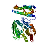
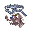
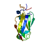
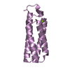
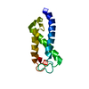
| Title | CRYSTAL STRUCTURE OF THE CATALYTIC DOMAIN OF YOPE-YERSINIA PESTIS GAP EFFECTOR PROTEIN. | ||||||
 Components Components | YERSINIA PESTIS VIRULENCE PROTEIN YOPE | ||||||
 Keywords Keywords | TOXIN / four helix up-down-up-down antiparallel bundle / beta hairpin / arginine finger | ||||||
| Function / homology |  Function and homology information Function and homology informationsymbiont-mediated perturbation of host actin cytoskeleton via filamentous actin depolymerization / negative regulation of phagocytosis / GTPase activator activity / cell outer membrane Similarity search - Function | ||||||
| Biological species |   Yersinia pestis (bacteria) Yersinia pestis (bacteria) | ||||||
| Method |  X-RAY DIFFRACTION / MAD / Resolution: 2.25 Å X-RAY DIFFRACTION / MAD / Resolution: 2.25 Å | ||||||
 Authors Authors | Evdokimov, A.G. / Tropea, J.E. / Routzahn, K.M. / Waugh, D.S. | ||||||
 Citation Citation | Journal: Protein Sci. / Year: 2002 Title: Crystal structure of the Yersinia pestis GTPase activator YopE. Authors: Evdokimov, A.G. / Tropea, J.E. / Routzahn, K.M. / Waugh, D.S. | ||||||
| History |
|
- Structure visualization
| Structure viewer | Molecule: MolmilJmol/JSmol |
|---|
- Downloads & links
-Download
| PDBx/mmCIF format | 1hy5.cif.gz | 60.3 KB | Display | PDBx/mmCIF format |
|---|---|---|---|---|
| PDB format | pdb1hy5.ent.gz | 43 KB | Display | PDB format |
| PDBx/mmJSON format | 1hy5.json.gz | Tree view | PDBx/mmJSON format | |
| Others |  Other downloads Other downloads |
-Validation report
| Summary document | 1hy5_validation.pdf.gz | 427.7 KB | Display | wwPDB validaton report |
|---|---|---|---|---|
| Full document | 1hy5_full_validation.pdf.gz | 434.5 KB | Display | |
| Data in XML | 1hy5_validation.xml.gz | 13.5 KB | Display | |
| Data in CIF | 1hy5_validation.cif.gz | 17.2 KB | Display | |
| Arichive directory | https://data.pdbj.org/pub/pdb/validation_reports/hy/1hy5ftp://data.pdbj.org/pub/pdb/validation_reports/hy/1hy5 | HTTPS FTP |
-Related structure data
| Related structure data | |
|---|---|
| Similar structure data |


-Links
 PDBj
PDBj- Assembly
| Deposited unit | 
| ||||||||
|---|---|---|---|---|---|---|---|---|---|
| 1 | 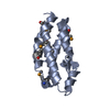
| ||||||||
| 2 | 
| ||||||||
| Unit cell |
| ||||||||
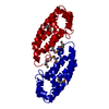
| Details | biological assembly is a monomer - crystal AU contains a dimer. RMSD of the Ca positions within the dimer is 0.34 A |
-Components
| #1: Protein | Mass: 14764.720 Da / Num. of mol.: 2 Fragment: C-TERMINAL DOMAIN (RESIDUES 90-219); BACTERIAL GTPASE ACTIVATING PROTEIN (GAP DOMAIN) Source method: isolated from a genetically manipulated source Source: (gene. exp.) Yersinia pestis (bacteria) / Gene: YOPE / Production host: #2: Water | ChemComp-HOH / |  Mass: 18.015 Da / Num. of mol.: 67 / Source method: isolated from a natural source / Formula: H2O Mass: 18.015 Da / Num. of mol.: 67 / Source method: isolated from a natural source / Formula: H2OHas protein modification | Y | |
|---|
-Experimental details
-Experiment
| Experiment | Method: X-RAY DIFFRACTION / Number of used crystals: 1 |
|---|
- Sample preparation
| Crystal | Density Matthews: 2.52 Å3/Da / Density % sol: 50.84 % | ||||||||||||||||||||||||
|---|---|---|---|---|---|---|---|---|---|---|---|---|---|---|---|---|---|---|---|---|---|---|---|---|---|
| Crystal grow | Temperature: 300 K / Method: vapor diffusion, hanging drop / pH: 9 Details: Ammonium sulphate, bicine, potassium nitrate, pH 9, VAPOR DIFFUSION, HANGING DROP, temperature 300K | ||||||||||||||||||||||||
| Crystal grow | *PLUS pH: 7 / Method: vapor diffusion | ||||||||||||||||||||||||
| Components of the solutions | *PLUS
|
-Data collection
| Diffraction | Mean temperature: 100 K |
|---|---|
| Diffraction source | Source: ROTATING ANODE / Type: RIGAKU / Wavelength: 1.5418 / Wavelength: 1.5418 Å |
| Detector | Type: MAR scanner 345 mm plate / Detector: IMAGE PLATE / Date: Oct 5, 2000 / Details: osmic mirrors |
| Radiation | Monochromator: graphite / Protocol: SINGLE WAVELENGTH / Monochromatic (M) / Laue (L): M / Scattering type: x-ray |
| Radiation wavelength | Wavelength: 1.5418 Å / Relative weight: 1 |
| Reflection | Resolution: 2.25→100 Å / Num. all: 22062 / Num. obs: 18478 / % possible obs: 96 % / Observed criterion σ(F): 0 / Observed criterion σ(I): 0 / Redundancy: 3.2 % / Biso Wilson estimate: 39 Å2 / Rmerge(I) obs: 0.04 / Rsym value: 0.06 / Net I/σ(I): 16 |
| Reflection shell | Resolution: 2.25→2.33 Å / Redundancy: 3 % / Rmerge(I) obs: 0.2 / Mean I/σ(I) obs: 4 / Num. unique all: 2291 / % possible all: 96.2 |
| Reflection | *PLUS Lowest resolution: 30 Å / Num. obs: 22062 / % possible obs: 98.9 % / Redundancy: 2.3 % / Rmerge(I) obs: 0.06 |
| Reflection shell | *PLUS % possible obs: 96.2 % / Redundancy: 1.8 % / Rmerge(I) obs: 0.221 / Mean I/σ(I) obs: 3.9 |
- Processing
| Software |
| |||||||||||||||||||||||||
|---|---|---|---|---|---|---|---|---|---|---|---|---|---|---|---|---|---|---|---|---|---|---|---|---|---|---|
| Refinement | Method to determine structure: MAD / Resolution: 2.25→100 Å Isotropic thermal model: isotropic individual atomic B factors Cross valid method: THROUGHOUT / σ(F): 0 / σ(I): 0 / Stereochemistry target values: Engh-Huber Details: Refinement on unmerged Friedel opposites (due to small but tangible selenium anomalous contribution). Refinement by conjugated-gradient least-squares. Selenomethionine restraints derived ...Details: Refinement on unmerged Friedel opposites (due to small but tangible selenium anomalous contribution). Refinement by conjugated-gradient least-squares. Selenomethionine restraints derived from Hic-Up. Two monomers were restrained by NCS (1,4-restraints generated by NCSY in SHELXL). Removal of the NCS restraints does not result in significant changes in the model. The structure was refined using home data (wavelength 1.5418).
| |||||||||||||||||||||||||
| Solvent computation | Solvent model: Babinet (SHELXL) / Bsol: 250.018 Å2 / ksol: 0.879 e/Å3 | |||||||||||||||||||||||||
| Displacement parameters | Biso mean: 33 Å2
| |||||||||||||||||||||||||
| Refine analyze | Luzzati coordinate error obs: 0.22 Å | |||||||||||||||||||||||||
| Refinement step | Cycle: LAST / Resolution: 2.25→100 Å
| |||||||||||||||||||||||||
| Refine LS restraints |
| |||||||||||||||||||||||||
| Software | *PLUS Name: SHELXL-97 / Classification: refinement | |||||||||||||||||||||||||
| Refinement | *PLUS Lowest resolution: 100 Å / σ(F): 0 / % reflection Rfree: 8 % | |||||||||||||||||||||||||
| Solvent computation | *PLUS | |||||||||||||||||||||||||
| Displacement parameters | *PLUS | |||||||||||||||||||||||||
| Refine LS restraints | *PLUS
|
