 Movie
Movie Controller

[English] 日本語
 Yorodumi
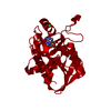
- PDB-1g4w: CRYSTAL STRUCTURE OF THE SALMONELLA TYROSINE PHOSPHATASE AND GTPA... -
+ Open data

- Basic information
| Entry | Database: PDB / ID: 1g4w | ||||||
|---|---|---|---|---|---|---|---|
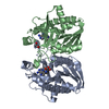
| Title | CRYSTAL STRUCTURE OF THE SALMONELLA TYROSINE PHOSPHATASE AND GTPASE ACTIVATING PROTEIN SPTP | ||||||
 Components Components | PROTEIN TYROSINE PHOSPHATASE SPTP | ||||||
 Keywords Keywords | SIGNALING PROTEIN / virulence factor / tyrosine phosphatase / GTPase activating protein / 4-helix bundle / disorder | ||||||
| Function / homology |  Function and homology information Function and homology informationprotein-tyrosine-phosphatase / protein tyrosine phosphatase activity / GTPase activator activity / host cell cytoplasm / : Similarity search - Function | ||||||
| Biological species |  Salmonella typhimurium (bacteria) Salmonella typhimurium (bacteria) | ||||||
| Method |  X-RAY DIFFRACTION / MIR / Resolution: 2.2 Å X-RAY DIFFRACTION / MIR / Resolution: 2.2 Å | ||||||
 Authors Authors | Stebbins, C.E. / Galan, J.E. | ||||||
 Citation Citation | Journal: Mol.Cell / Year: 2000 Title: Modulation of host signaling by a bacterial mimic: structure of the Salmonella effector SptP bound to Rac1. Authors: Stebbins, C.E. / Galan, J.E. | ||||||
| History |
|
- Structure visualization
| Structure viewer | Molecule: MolmilJmol/JSmol |
|---|
- Downloads & links
-Download
| PDBx/mmCIF format | 1g4w.cif.gz | 80.5 KB | Display | PDBx/mmCIF format |
|---|---|---|---|---|
| PDB format | pdb1g4w.ent.gz | 60.6 KB | Display | PDB format |
| PDBx/mmJSON format | 1g4w.json.gz | Tree view | PDBx/mmJSON format | |
| Others |  Other downloads Other downloads |
-Validation report
| Arichive directory | https://data.pdbj.org/pub/pdb/validation_reports/g4/1g4wftp://data.pdbj.org/pub/pdb/validation_reports/g4/1g4w | HTTPS FTP |
|---|
-Related structure data


-Links
 PDBj
PDBj


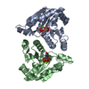
- Assembly
| Deposited unit | 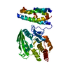
| ||||||||
|---|---|---|---|---|---|---|---|---|---|
| 1 |
| ||||||||
| Unit cell |
|
-Components
| #1: Protein | Mass: 42127.801 Da / Num. of mol.: 1 / Fragment: SPTP RESIDUES 161-543 Source method: isolated from a genetically manipulated source Source: (gene. exp.) Salmonella typhimurium (bacteria) / Gene: SPTP / Plasmid: PGEX-4T-3 / Species (production host): Escherichia coli / Production host: |
|---|---|
| #2: Water | ChemComp-HOH /  Mass: 18.015 Da / Num. of mol.: 154 / Source method: isolated from a natural source / Formula: H2O Mass: 18.015 Da / Num. of mol.: 154 / Source method: isolated from a natural source / Formula: H2O |
-Experimental details
-Experiment
| Experiment | Method: X-RAY DIFFRACTION / Number of used crystals: 1 |
|---|
- Sample preparation
| Crystal | Density Matthews: 2.79 Å3/Da / Density % sol: 55.99 % | ||||||||||||||||||||
|---|---|---|---|---|---|---|---|---|---|---|---|---|---|---|---|---|---|---|---|---|---|
| Crystal grow | Temperature: 295 K / Method: vapor diffusion, hanging drop / pH: 8 Details: 3.5M sodium formate, 2mM DTT, 0.1M Tris pH 8.0, VAPOR DIFFUSION, HANGING DROP, temperature 295K | ||||||||||||||||||||
| Crystal | *PLUS Density % sol: 52 % | ||||||||||||||||||||
| Crystal grow | *PLUS | ||||||||||||||||||||
| Components of the solutions | *PLUS
|
-Data collection
| Diffraction | Mean temperature: 113 K |
|---|---|
| Diffraction source | Source: ROTATING ANODE / Type: RIGAKU RU200 / Wavelength: 1.5418 |
| Detector | Type: RIGAKU RAXIS IV / Detector: IMAGE PLATE / Date: Sep 21, 1999 / Details: Yale mirrors |
| Radiation | Monochromator: Yale Mirrors / Protocol: SINGLE WAVELENGTH / Monochromatic (M) / Laue (L): M / Scattering type: x-ray |
| Radiation wavelength | Wavelength: 1.5418 Å / Relative weight: 1 |
| Reflection | Resolution: 2.2→50 Å / Num. all: 24740 / Num. obs: 23440 / % possible obs: 94.8 % / Observed criterion σ(F): 1 / Observed criterion σ(I): 1 / Redundancy: 4.8 % / Biso Wilson estimate: 21.7 Å2 / Rmerge(I) obs: 0.077 / Net I/σ(I): 13.9 |
| Reflection shell | Resolution: 2.2→2.3 Å / Redundancy: 2 % / Rmerge(I) obs: 0.337 / Mean I/σ(I) obs: 2.93 / % possible all: 94.9 |
| Reflection | *PLUS Highest resolution: 2.2 Å / Num. measured all: 114569 |
- Processing
| Software |
| ||||||||||||||||||||
|---|---|---|---|---|---|---|---|---|---|---|---|---|---|---|---|---|---|---|---|---|---|
| Refinement | Method to determine structure: MIR / Resolution: 2.2→50 Å / σ(F): 0 / σ(I): 0 / Stereochemistry target values: Engh & Huber
| ||||||||||||||||||||
| Refinement step | Cycle: LAST / Resolution: 2.2→50 Å
| ||||||||||||||||||||
| Refine LS restraints |
| ||||||||||||||||||||
| LS refinement shell | Resolution: 2.2→2.23 Å /
| ||||||||||||||||||||
| Software | *PLUS Name: CNS / Classification: refinement | ||||||||||||||||||||
| Refinement | *PLUS Highest resolution: 2.2 Å / Lowest resolution: 50 Å / σ(F): 0 / % reflection Rfree: 7 % / Rfactor obs: 0.26 / Rfactor Rwork: 0.26 | ||||||||||||||||||||
| Solvent computation | *PLUS | ||||||||||||||||||||
| Displacement parameters | *PLUS | ||||||||||||||||||||
| Refine LS restraints | *PLUS Type: c_angle_deg / Dev ideal: 1.4 | ||||||||||||||||||||
| LS refinement shell | *PLUS Rfactor Rfree: 0.392 / Rfactor Rwork: 0.381 |
