 Movie
Movie Controller


+ Open data

- Basic information
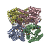
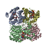
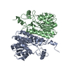
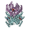
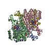
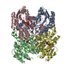
| Entry | Database: PDB / ID: 1hxh | ||||||
|---|---|---|---|---|---|---|---|
| Title | COMAMONAS TESTOSTERONI 3BETA/17BETA HYDROXYSTEROID DEHYDROGENASE | ||||||
 Components Components | 3BETA/17BETA-HYDROXYSTEROID DEHYDROGENASE | ||||||
 Keywords Keywords | OXIDOREDUCTASE / alpha-beta / Rossmann fold / short-chain dehydrogenase | ||||||
| Function / homology |  Function and homology information Function and homology information3(or 17)beta-hydroxysteroid dehydrogenase / testosterone dehydrogenase (NADP+) activity / testosterone dehydrogenase (NAD+) activity / steroid metabolic process Similarity search - Function | ||||||
| Biological species |  Comamonas testosteroni (bacteria) Comamonas testosteroni (bacteria) | ||||||
| Method |  X-RAY DIFFRACTION / SYNCHROTRON / MOLECULAR REPLACEMENT / Resolution: 1.22 Å X-RAY DIFFRACTION / SYNCHROTRON / MOLECULAR REPLACEMENT / Resolution: 1.22 Å | ||||||
 Authors Authors | Benach, J. / Filling, C. / Oppermann, U.C.T. / Roversi, P. / Bricogne, G. / Berndt, K.D. / Jornvall, H. / Ladenstein, R. | ||||||
 Citation Citation | Journal: Biochemistry / Year: 2002 Title: Structure of Bacterial 3beta/17beta-Hydroxysteroid Dehydrogenase at 1.2 A Resolution: A Model for Multiple Steroid Recognition Authors: Benach, J. / Filling, C. / Oppermann, U.C.T. / Roversi, P. / Bricogne, G. / Berndt, K.D. / Jornvall, H. / Ladenstein, R. | ||||||
| History |
|
- Structure visualization
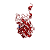
| Structure viewer | Molecule: MolmilJmol/JSmol |
|---|
- Downloads & links
-Download
| PDBx/mmCIF format | 1hxh.cif.gz | 414 KB | Display | PDBx/mmCIF format |
|---|---|---|---|---|
| PDB format | pdb1hxh.ent.gz | 339.2 KB | Display | PDB format |
| PDBx/mmJSON format | 1hxh.json.gz | Tree view | PDBx/mmJSON format | |
| Others |  Other downloads Other downloads |
-Validation report
| Arichive directory | https://data.pdbj.org/pub/pdb/validation_reports/hx/1hxhftp://data.pdbj.org/pub/pdb/validation_reports/hx/1hxh | HTTPS FTP |
|---|
-Related structure data
| Related structure data |  2hsdS S: Starting model for refinement |
|---|---|
| Similar structure data |


-Links
 PDBj
PDBj


- Assembly
| Deposited unit | 
| ||||||||
|---|---|---|---|---|---|---|---|---|---|
| 1 |
| ||||||||
| Unit cell |
|
-Components
| #1: Protein | Mass: 26847.855 Da / Num. of mol.: 4 Source method: isolated from a genetically manipulated source Source: (gene. exp.) Comamonas testosteroni (bacteria) / Plasmid: PET15B / Species (production host): Escherichia coli / Production host: References: UniProt: P19871, 3(or 17)beta-hydroxysteroid dehydrogenase #2: Water | ChemComp-HOH / |  Mass: 18.015 Da / Num. of mol.: 1256 / Source method: isolated from a natural source / Formula: H2O Mass: 18.015 Da / Num. of mol.: 1256 / Source method: isolated from a natural source / Formula: H2O |
|---|
-Experimental details
-Experiment
| Experiment | Method: X-RAY DIFFRACTION / Number of used crystals: 1 |
|---|
- Sample preparation
| Crystal | Density Matthews: 2.36 Å3/Da / Density % sol: 47.58 % | ||||||||||||||||||||||||||||||||||||||||||
|---|---|---|---|---|---|---|---|---|---|---|---|---|---|---|---|---|---|---|---|---|---|---|---|---|---|---|---|---|---|---|---|---|---|---|---|---|---|---|---|---|---|---|---|
| Crystal grow | Temperature: 277 K / Method: vapor diffusion, sitting drop / pH: 7.5 Details: 10% Ethanol, 250 mM NaCl, 50 mM Tris/HCl, 10 mM DTT, pH 7.5, VAPOR DIFFUSION, SITTING DROP, temperature 277K | ||||||||||||||||||||||||||||||||||||||||||
| Crystal grow | *PLUS Method: vapor diffusion | ||||||||||||||||||||||||||||||||||||||||||
| Components of the solutions | *PLUS
|
-Data collection
| Diffraction | Mean temperature: 100 K |
|---|---|
| Diffraction source | Source: SYNCHROTRON / Site: EMBL/DESY, HAMBURG  / Beamline: BW7B / Wavelength: 0.8424 Å / Beamline: BW7B / Wavelength: 0.8424 Å |
| Detector | Type: MARRESEARCH / Detector: IMAGE PLATE / Date: Mar 15, 2000 |
| Radiation | Protocol: SINGLE WAVELENGTH / Monochromatic (M) / Laue (L): M / Scattering type: x-ray |
| Radiation wavelength | Wavelength: 0.8424 Å / Relative weight: 1 |
| Reflection | Resolution: 1.18→40 Å / Num. all: 1409412 / Num. obs: 1409412 / % possible obs: 94.3 % / Observed criterion σ(F): 2 / Observed criterion σ(I): -1 / Redundancy: 4 % / Biso Wilson estimate: 11 Å2 / Rmerge(I) obs: 0.069 |
| Reflection shell | Resolution: 1.22→1.25 Å / Rmerge(I) obs: 0.383 / % possible all: 83.9 |
| Reflection | *PLUS Lowest resolution: 40 Å / Num. obs: 314581 / Num. measured all: 1409412 |
| Reflection shell | *PLUS % possible obs: 83.9 % / Mean I/σ(I) obs: 54.6 |
- Processing
| Software |
| |||||||||||||||||||||
|---|---|---|---|---|---|---|---|---|---|---|---|---|---|---|---|---|---|---|---|---|---|---|
| Refinement | Method to determine structure: MOLECULAR REPLACEMENT Starting model: 2HSD Resolution: 1.22→20 Å / SU ML: 0.15 / Cross valid method: free / σ(F): 0 / σ(I): 0 / ESU R: 0.16 / ESU R Free: 0.17
| |||||||||||||||||||||
| Displacement parameters | Biso mean: 19 Å2 | |||||||||||||||||||||
| Refinement step | Cycle: LAST / Resolution: 1.22→20 Å
| |||||||||||||||||||||
| Refine LS restraints |
| |||||||||||||||||||||
| LS refinement shell | Resolution: 1.22→20 Å / Rfactor Rfree: 0.18 / Rfactor Rwork: 0.146 | |||||||||||||||||||||
| Refinement | *PLUS Lowest resolution: 20 Å / Rfactor Rfree: 0.18 | |||||||||||||||||||||
| Solvent computation | *PLUS | |||||||||||||||||||||
| Displacement parameters | *PLUS | |||||||||||||||||||||
| Refine LS restraints | *PLUS
| |||||||||||||||||||||
| LS refinement shell | *PLUS Rfactor Rfree: 0.172 / Rfactor Rwork: 0.139 |
