 Movie
Movie Controller


+ Open data

- Basic information
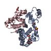
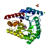
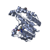
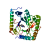
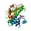
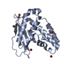
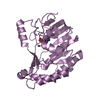
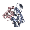
| Entry | Database: PDB / ID: 1hw6 | ||||||
|---|---|---|---|---|---|---|---|
| Title | CRYSTAL STRUCTURE OF APO-2,5-DIKETO-D-GLUCONATE REDUCTASE | ||||||
 Components Components | 2,5-DIKETO-D-GLUCONIC ACID REDUCTASE | ||||||
 Keywords Keywords | OXIDOREDUCTASE / aldo-keto reductase / TIM barrel | ||||||
| Function / homology |  Function and homology information Function and homology information2,5-didehydrogluconate reductase (2-dehydro-L-gulonate-forming) / L-ascorbic acid biosynthetic process / : / cytoplasm Similarity search - Function | ||||||
| Biological species |  Corynebacterium sp. (bacteria) Corynebacterium sp. (bacteria) | ||||||
| Method |  X-RAY DIFFRACTION / MOLECULAR REPLACEMENT / Resolution: 1.9 Å X-RAY DIFFRACTION / MOLECULAR REPLACEMENT / Resolution: 1.9 Å | ||||||
 Authors Authors | Sanli, G. / Blaber, M. | ||||||
 Citation Citation | Journal: J.Mol.Biol. / Year: 2001 Title: Structural assembly of the active site in an aldo-keto reductase by NADPH cofactor. Authors: Sanli, G. / Blaber, M. | ||||||
| History |
|
- Structure visualization
| Structure viewer | Molecule: MolmilJmol/JSmol |
|---|
- Downloads & links
-Download
| PDBx/mmCIF format | 1hw6.cif.gz | 69.2 KB | Display | PDBx/mmCIF format |
|---|---|---|---|---|
| PDB format | pdb1hw6.ent.gz | 48.7 KB | Display | PDB format |
| PDBx/mmJSON format | 1hw6.json.gz | Tree view | PDBx/mmJSON format | |
| Others |  Other downloads Other downloads |
-Validation report
| Summary document | 1hw6_validation.pdf.gz | 418.2 KB | Display | wwPDB validaton report |
|---|---|---|---|---|
| Full document | 1hw6_full_validation.pdf.gz | 427 KB | Display | |
| Data in XML | 1hw6_validation.xml.gz | 15.3 KB | Display | |
| Data in CIF | 1hw6_validation.cif.gz | 22.5 KB | Display | |
| Arichive directory | https://data.pdbj.org/pub/pdb/validation_reports/hw/1hw6ftp://data.pdbj.org/pub/pdb/validation_reports/hw/1hw6 | HTTPS FTP |
-Related structure data
| Related structure data |  1a80S S: Starting model for refinement |
|---|---|
| Similar structure data |


-Links
 PDBj
PDBj
- Assembly
| Deposited unit | 
| ||||||||
|---|---|---|---|---|---|---|---|---|---|
| 1 |
| ||||||||
| Unit cell |
|
-Components
| #1: Protein | Mass: 30151.664 Da / Num. of mol.: 1 Source method: isolated from a genetically manipulated source Source: (gene. exp.) Corynebacterium sp. (bacteria) / Production host:   Spodoptera frugiperda (fall armyworm) Spodoptera frugiperda (fall armyworm)References: UniProt: P06632, Oxidoreductases; Acting on the CH-OH group of donors; With NAD+ or NADP+ as acceptor | ||||
|---|---|---|---|---|---|
| #2: Chemical |   Mass: 24.305 Da / Num. of mol.: 2 / Source method: obtained synthetically / Formula: Mg Mass: 24.305 Da / Num. of mol.: 2 / Source method: obtained synthetically / Formula: Mg#3: Chemical |   Mass: 35.453 Da / Num. of mol.: 2 / Source method: obtained synthetically / Formula: Cl Mass: 35.453 Da / Num. of mol.: 2 / Source method: obtained synthetically / Formula: Cl#4: Water | ChemComp-HOH / |  Mass: 18.015 Da / Num. of mol.: 265 / Source method: isolated from a natural source / Formula: H2O Mass: 18.015 Da / Num. of mol.: 265 / Source method: isolated from a natural source / Formula: H2O |
-Experimental details
-Experiment
| Experiment | Method: X-RAY DIFFRACTION / Number of used crystals: 1 |
|---|
- Sample preparation
| Crystal | Density Matthews: 2.2 Å3/Da / Density % sol: 42.18 % | ||||||||||||||||||||||||||||||||||||
|---|---|---|---|---|---|---|---|---|---|---|---|---|---|---|---|---|---|---|---|---|---|---|---|---|---|---|---|---|---|---|---|---|---|---|---|---|---|
| Crystal grow | Temperature: 298 K / Method: vapor diffusion, hanging drop / pH: 8.5 Details: 30% PEG 4000, 0.2M MgCl2, 0.1M Tris, pH 8.5, VAPOR DIFFUSION, HANGING DROP, temperature 298K | ||||||||||||||||||||||||||||||||||||
| Crystal grow | *PLUS pH: 7.5 | ||||||||||||||||||||||||||||||||||||
| Components of the solutions | *PLUS
|
-Data collection
| Diffraction | Mean temperature: 103 K |
|---|---|
| Diffraction source | Source: ROTATING ANODE / Type: RIGAKU / Wavelength: 1.5418 Å |
| Detector | Type: RIGAKU RAXIS II / Detector: IMAGE PLATE / Date: Jan 1, 2000 / Details: mirrors |
| Radiation | Monochromator: Osmic mirrors / Protocol: SINGLE WAVELENGTH / Monochromatic (M) / Laue (L): M / Scattering type: x-ray |
| Radiation wavelength | Wavelength: 1.5418 Å / Relative weight: 1 |
| Reflection | Resolution: 1.9→27 Å / Num. all: 19905 / Num. obs: 126729 / % possible obs: 94.5 % / Observed criterion σ(I): 3 / Redundancy: 6.4 % / Biso Wilson estimate: 16.1 Å2 / Rmerge(I) obs: 0.052 / Net I/σ(I): 15.6 |
| Reflection shell | Resolution: 1.9→1.94 Å / Redundancy: 2.24 % / Rmerge(I) obs: 0.343 / Mean I/σ(I) obs: 3.2 / % possible all: 82.8 |
| Reflection | *PLUS Num. obs: 19905 / Num. measured all: 126729 |
| Reflection shell | *PLUS Highest resolution: 1.9 Å / % possible obs: 82.8 % |
- Processing
| Software |
| |||||||||||||||||||||
|---|---|---|---|---|---|---|---|---|---|---|---|---|---|---|---|---|---|---|---|---|---|---|
| Refinement | Method to determine structure: MOLECULAR REPLACEMENT Starting model: 1A80 Resolution: 1.9→27 Å / Isotropic thermal model: TRONRUD / Stereochemistry target values: TRONRUD
| |||||||||||||||||||||
| Refinement step | Cycle: LAST / Resolution: 1.9→27 Å
| |||||||||||||||||||||
| Refine LS restraints |
| |||||||||||||||||||||
| LS refinement shell | Resolution: 1.9→1.93 Å
| |||||||||||||||||||||
| Software | *PLUS Name: TNT / Version: 5E / Classification: refinement | |||||||||||||||||||||
| Refinement | *PLUS Rfactor obs: 0.2 / Rfactor Rfree: 0.268 / Rfactor Rwork: 0.2 | |||||||||||||||||||||
| Solvent computation | *PLUS | |||||||||||||||||||||
| Displacement parameters | *PLUS | |||||||||||||||||||||
| Refine LS restraints | *PLUS
|
