 Movie
Movie Controller

[English] 日本語
 Yorodumi
- PDB-1ht1: Nucleotide-Dependent Conformational Changes in a Protease-Associa... -
+ Open data

- Basic information
| Entry | Database: PDB / ID: 1ht1 | ||||||
|---|---|---|---|---|---|---|---|
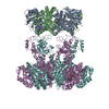
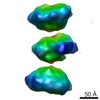
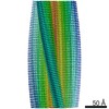
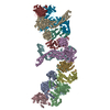
| Title | Nucleotide-Dependent Conformational Changes in a Protease-Associated ATPase HslU | ||||||
 Components Components |
| ||||||
 Keywords Keywords | CHAPERONE / HSLVU / PEPTIDASE-ATPASE COMPLEX | ||||||
| Function / homology |  Function and homology information Function and homology informationprotein denaturation / HslU-HslV peptidase / HslUV protease complex / proteasome-activating activity / proteasome core complex / protein unfolding / threonine-type endopeptidase activity / : / peptidase activity / cellular response to heat ...protein denaturation / HslU-HslV peptidase / HslUV protease complex / proteasome-activating activity / proteasome core complex / protein unfolding / threonine-type endopeptidase activity / : / peptidase activity / cellular response to heat / response to heat / protein domain specific binding / magnesium ion binding / ATP hydrolysis activity / proteolysis / ATP binding / membrane / identical protein binding / cytoplasm / cytosol Similarity search - Function | ||||||
| Biological species |  | ||||||
| Method |  X-RAY DIFFRACTION / Resolution: 2.8 Å X-RAY DIFFRACTION / Resolution: 2.8 Å | ||||||
 Authors Authors | Wang, J. / Song, J.J. / Seong, I.S. / Franklin, M.C. / Kamtekar, S. / Eom, S.H. / Chung, C.H. | ||||||
 Citation Citation | Journal: Structure / Year: 2001 Title: Nucleotide-dependent conformational changes in a protease-associated ATPase HsIU. Authors: Wang, J. / Song, J.J. / Seong, I.S. / Franklin, M.C. / Kamtekar, S. / Eom, S.H. / Chung, C.H. #1: Journal: Proc.Natl.Acad.Sci.USA / Year: 2000Title: Mutational Studies of Hslu and its Docking Mode With Hslv. Authors: Song, H.K. / Hartmann, C. / Ramachandran, R. / Bochtler, M. / Behrendt, R. / Moroder, L. / Huber, R. #2: Journal: Nature / Year: 2000Title: The Structures of Hslu and the ATP-Dependent Protease HslU-HslV. Authors: Bochtler, M. / Hartmann, C. / Song, H.K. / Bourenkov, G.P. / Bartunik, H.D. / Huber, R. #3: Journal: Cell(Cambridge,Mass.) / Year: 2000Title: Crystal and Solution Structures of an HslUV Protease-chaperone Complex. Authors: Sousa, M.C. / Trame, C.B. / Tsuruta, S. / Wilbanks, S.M. / Reddy, V.S. / McKay, D.B. #4: Journal: Structure / Year: 2001Title: Crystal Structures of the Hslvu Peptidase-ATPase Complex Reveal an ATP-Dependent Proteolysis Mechanism Authors: Wang, J. / Song, J.J. / Franklin, M.C. / Kamtekar, S. / Im, Y.J. / Rho, S.H. / Seong, I.S. / Lee, C.S. / Chung, C.H. / Eom, S.H. #5: Journal: Nature / Year: 2000Title: ATP-dependent proteases: Docking of Components in a Bacterial Complex Authors: Ishikawa, T. / Maurizi, M.R. / Belnap, D. / Steven, A.C. #6: Journal: J.STRUCT.BIOL. / Year: 2001Title: A corrected quaternary arrangement of the peptidase hslv and atpase hslu in a cocrystal structure Authors: Wang, J. | ||||||
| History |
|
- Structure visualization
| Structure viewer | Molecule: MolmilJmol/JSmol |
|---|
- Downloads & links
-Download
| PDBx/mmCIF format | 1ht1.cif.gz | 574.2 KB | Display | PDBx/mmCIF format |
|---|---|---|---|---|
| PDB format | pdb1ht1.ent.gz | 471.3 KB | Display | PDB format |
| PDBx/mmJSON format | 1ht1.json.gz | Tree view | PDBx/mmJSON format | |
| Others |  Other downloads Other downloads |
-Validation report
| Arichive directory | https://data.pdbj.org/pub/pdb/validation_reports/ht/1ht1ftp://data.pdbj.org/pub/pdb/validation_reports/ht/1ht1 | HTTPS FTP |
|---|
-Related structure data


-Links
 PDBj
PDBj


- Assembly
| Deposited unit | 
| ||||||||
|---|---|---|---|---|---|---|---|---|---|
| 1 | 
| ||||||||
| 2 | 
| ||||||||
| Unit cell |
|
-Components
| #1: Protein | Mass: 18986.641 Da / Num. of mol.: 8 Source method: isolated from a genetically manipulated source Source: (gene. exp.) #2: Protein | Mass: 50495.531 Da / Num. of mol.: 4 Source method: isolated from a genetically manipulated source Source: (gene. exp.) #3: Chemical | ChemComp-ADP /   Mass: 427.201 Da / Num. of mol.: 4 / Source method: obtained synthetically / Formula: C10H15N5O10P2 / Comment: ADP, energy-carrying molecule*YM Mass: 427.201 Da / Num. of mol.: 4 / Source method: obtained synthetically / Formula: C10H15N5O10P2 / Comment: ADP, energy-carrying molecule*YM |
|---|
-Experimental details
-Experiment
| Experiment | Method: X-RAY DIFFRACTION |
|---|
- Sample preparation
| Crystal | Density Matthews: 3.34 Å3/Da / Density % sol: 63.14 % |
|---|---|
| Crystal grow | *PLUS Method: unknown |
-Data collection
| Radiation | Protocol: SINGLE WAVELENGTH / Monochromatic (M) / Laue (L): M / Scattering type: x-ray |
|---|---|
| Radiation wavelength | Relative weight: 1 |
- Processing
| Software | Name: CNS / Version: 1 / Classification: refinement | ||||||||||||||||||||
|---|---|---|---|---|---|---|---|---|---|---|---|---|---|---|---|---|---|---|---|---|---|
| Refinement | Resolution: 2.8→29.62 Å / Data cutoff high absF: 683901.76 / Data cutoff low absF: 0 / Cross valid method: THROUGHOUT / σ(F): 0 Details: THIS ENTRY CONTAINS A SOLUTION IN X-RAY STRUCTURES TO THE X-RAY DIFFRACTION DATA THAT WERE RETRIEVED FROM PDB DATABASE UNDER ACCESSION NUMBER 1E94. THIS ENTRY IS RELATED TO 1HQY, 1HT2 AND ...Details: THIS ENTRY CONTAINS A SOLUTION IN X-RAY STRUCTURES TO THE X-RAY DIFFRACTION DATA THAT WERE RETRIEVED FROM PDB DATABASE UNDER ACCESSION NUMBER 1E94. THIS ENTRY IS RELATED TO 1HQY, 1HT2 AND 1E94. THE ASSIGNMENT OF THE SCREW AXIS 6(3) IN THE P6(3)22 SPACE GROUP FOR THE HSLVU COMPLEX STRUCTURES DESCRIBED IN REFERENCES 1 AND 2 (CORRESPONDING PDB ACCESSION NUMBERS ARE 1DOO AND 1E94, RESPECTIVELY) REQUIRES THE PRESENCE OF SYSTEMATIC EXTINCTIONS ALONG (00L) WITH L=2N+1. THERE WERE NO SYSTEMATIC EXTINCTIONS AT ALL IN THE 1E94SF ENTRY. THERE WERE TWO REFLECTIONS WITH F/SIGMA(F) NEAR 20 AND NINE REFLECTIONS WITH F/SIMGA(F) OVER 10 ALONG (00L) WITH L=2N+1. SUCH A LARGE NUMBER OF SIGNIFICANT OBSERVATIONS CANNOT BE DUE TO TECHNICAL ERRORS IN MEASUREMENT OF X-RAY DIFFRACTION DATA. A STATISTICAL ANALYSIS OF THEM COMPARED WITH THE REST OF THE DATA CONFIRMS THAT THEY ARE NOT DUE TO TECHNICAL ERRORS. THEREFORE, THE SPACE GROUP MUST NOT BE P6(3)22. LARGE VALUES OF COMBINED R-MERGE VALUES FOR OBSERVED DATA 1DOO (14.1%) AND 1E94 (12.1%) ARE INDICATIVE OF INCORRECT ASSIGNEMENT OF POINT SYMMETRY GROUP TO BE 622. THE ESTIMATED MEASUREMENT PRECISION IN INTENSITY SHOULD BE ABOUT 1% ON THE BASIS OF THE AVERAGE F/SIGMA(F) OF 44.7 IN 1E94 OBSERVED DATA. THEREFORE, ONE SETS OF DYADS IN THE POINT SYMMETRY P622 WERE TWINNING OPERATIONS. THERE WERE LARGE DISCREPANCIES IN WILSON RATIO (
| ||||||||||||||||||||
| Refinement step | Cycle: LAST / Resolution: 2.8→29.62 Å
| ||||||||||||||||||||
| Xplor file |
|
