 Movie
Movie Controller

[English] 日本語
 Yorodumi
- PDB-1hst: CRYSTAL STRUCTURE OF GLOBULAR DOMAIN OF HISTONE H5 AND ITS IMPLIC... -
+ Open data

- Basic information
| Entry | Database: PDB / ID: 1hst | ||||||
|---|---|---|---|---|---|---|---|
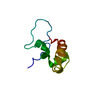
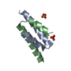
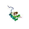
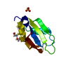
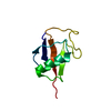
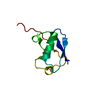
| Title | CRYSTAL STRUCTURE OF GLOBULAR DOMAIN OF HISTONE H5 AND ITS IMPLICATIONS FOR NUCLEOSOME BINDING | ||||||
 Components Components | HISTONE H5 | ||||||
 Keywords Keywords | CHROMOSOMAL PROTEIN | ||||||
| Function / homology |  Function and homology information Function and homology informationnegative regulation of DNA recombination / chromosome condensation / nucleosomal DNA binding / structural constituent of chromatin / nucleosome / nucleosome assembly / chromatin organization / double-stranded DNA binding / nucleus Similarity search - Function | ||||||
| Biological species |  | ||||||
| Method |  X-RAY DIFFRACTION / Resolution: 2.6 Å X-RAY DIFFRACTION / Resolution: 2.6 Å | ||||||
 Authors Authors | Ramakrishnan, V. / Finch, J.T. / Graziano, V. / Lee, P.L. / Sweet, R.M. | ||||||
 Citation Citation | Journal: Nature / Year: 1993 Title: Crystal structure of globular domain of histone H5 and its implications for nucleosome binding. Authors: Ramakrishnan, V. / Finch, J.T. / Graziano, V. / Lee, P.L. / Sweet, R.M. #1: Journal: J.Mol.Biol. / Year: 1990Title: Crystallization of the Globular Domain of Histone H5 Authors: Graziano, V. / Gerchman, S.E. / Wonacott, A.J. / Sweet, R.M. / Wells, J.R.E. / White, S.W. / Ramakrishnan, V. | ||||||
| History |
|
- Structure visualization
| Structure viewer | Molecule: MolmilJmol/JSmol |
|---|
- Downloads & links
-Download
| PDBx/mmCIF format | 1hst.cif.gz | 38.5 KB | Display | PDBx/mmCIF format |
|---|---|---|---|---|
| PDB format | pdb1hst.ent.gz | 27.3 KB | Display | PDB format |
| PDBx/mmJSON format | 1hst.json.gz | Tree view | PDBx/mmJSON format | |
| Others |  Other downloads Other downloads |
-Validation report
| Summary document | 1hst_validation.pdf.gz | 374.8 KB | Display | wwPDB validaton report |
|---|---|---|---|---|
| Full document | 1hst_full_validation.pdf.gz | 379.9 KB | Display | |
| Data in XML | 1hst_validation.xml.gz | 5 KB | Display | |
| Data in CIF | 1hst_validation.cif.gz | 6.9 KB | Display | |
| Arichive directory | https://data.pdbj.org/pub/pdb/validation_reports/hs/1hstftp://data.pdbj.org/pub/pdb/validation_reports/hs/1hst | HTTPS FTP |
-Related structure data
| Similar structure data |
|---|


-Links
 PDBj
PDBj- Assembly
| Deposited unit | 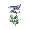
| ||||||||
|---|---|---|---|---|---|---|---|---|---|
| 1 | 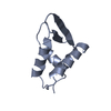
| ||||||||
| 2 | 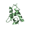
| ||||||||
| Unit cell |
| ||||||||
| Atom site foot note | 1: RESIDUES A 39 - A 44 AND B 39 - B 44 FORM A LOOP BETWEEN HELICES I AND II. THESE ARE NOT WELL DEFINED IN THE DENSITY, AND THEIR POSITIONS ARE ONLY PRELIMINARY. | ||||||||
| Noncrystallographic symmetry (NCS) | NCS oper: (Code: given Matrix: (-0.7827, -0.2417, 0.5735), Vector: Details | THE TRANSFORMATION PRESENTED ON *MTRIX* RECORDS BELOW WILL YIELD APPROXIMATE COORDINATES FOR CHAIN *B* WHEN APPLIED TO CHAIN *A*. HOWEVER, THE CONFORMATION OF THE REGION FROM RESIDUE 78 TO RESIDUE 97 IS DIFFERENT IN THE TWO CHAINS. | |
-Components
| #1: Protein | Mass: 9789.352 Da / Num. of mol.: 2 Source method: isolated from a genetically manipulated source Source: (gene. exp.) |
|---|
-Experimental details
-Experiment
| Experiment | Method: X-RAY DIFFRACTION |
|---|
- Sample preparation
| Crystal | Density Matthews: 2.74 Å3/Da / Density % sol: 55.03 % |
|---|---|
| Crystal grow | *PLUS pH: 8.2 / Method: vapor diffusion, hanging drop / Details: referred to J.Mol.Biol. 212.253-257 1990 |
| Components of the solutions | *PLUS Conc.: 2.2 M / Common name: phosphate |
-Data collection
| Radiation | Scattering type: x-ray |
|---|---|
| Radiation wavelength | Relative weight: 1 |
- Processing
| Software |
| ||||||||||||||||||||||||||||||||||||||||||||||||||||||||||||
|---|---|---|---|---|---|---|---|---|---|---|---|---|---|---|---|---|---|---|---|---|---|---|---|---|---|---|---|---|---|---|---|---|---|---|---|---|---|---|---|---|---|---|---|---|---|---|---|---|---|---|---|---|---|---|---|---|---|---|---|---|---|
| Refinement | Resolution: 2.6→6 Å / Rfactor Rwork: 0.27 / Rfactor obs: 0.27 / σ(F): 0 Details: RESIDUES 24 - 96 ARE REPRESENTED IN THIS ENTRY, ALTHOUGH POORLY DEFINED DENSITY IS VISIBLE BEYOND RESIDUE 96 OF CHAIN *A*. OF THE 89 RESIDUES OF THE POLYPEPTIDE CHAIN, 5 RESIDUES AT THE N- ...Details: RESIDUES 24 - 96 ARE REPRESENTED IN THIS ENTRY, ALTHOUGH POORLY DEFINED DENSITY IS VISIBLE BEYOND RESIDUE 96 OF CHAIN *A*. OF THE 89 RESIDUES OF THE POLYPEPTIDE CHAIN, 5 RESIDUES AT THE N-TERMINUS AND 11 RESIDUES AT THE C-TERMINUS HAVE NOT BEEN INCLUDED IN THE MODEL; THUS THE R-FACTOR IS SOMEWHAT HIGH AT THIS POINT. | ||||||||||||||||||||||||||||||||||||||||||||||||||||||||||||
| Refinement step | Cycle: LAST / Resolution: 2.6→6 Å
| ||||||||||||||||||||||||||||||||||||||||||||||||||||||||||||
| Refine LS restraints |
| ||||||||||||||||||||||||||||||||||||||||||||||||||||||||||||
| Refinement | *PLUS Highest resolution: 2.6 Å / Lowest resolution: 6 Å / σ(F): 0 / Rfactor all: 0.27 | ||||||||||||||||||||||||||||||||||||||||||||||||||||||||||||
| Solvent computation | *PLUS | ||||||||||||||||||||||||||||||||||||||||||||||||||||||||||||
| Displacement parameters | *PLUS | ||||||||||||||||||||||||||||||||||||||||||||||||||||||||||||
| Refine LS restraints | *PLUS Type: x_angle_d / Dev ideal: 3.1 |
