 Movie
Movie Controller

[English] 日本語
 Yorodumi
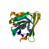
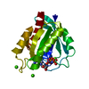
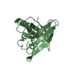
- PDB-1hqp: CRYSTAL STRUCTURE OF A TRUNCATED FORM OF PORCINE ODORANT-BINDING ... -
+ Open data

- Basic information
| Entry | Database: PDB / ID: 1hqp | ||||||
|---|---|---|---|---|---|---|---|
| Title | CRYSTAL STRUCTURE OF A TRUNCATED FORM OF PORCINE ODORANT-BINDING PROTEIN | ||||||
 Components Components | ODORANT-BINDING PROTEIN | ||||||
 Keywords Keywords | SIGNALING PROTEIN / lipocalin / dimer / ligand-binding site / access to the binding site | ||||||
| Function / homology |  Function and homology information Function and homology informationodorant binding / small molecule binding / sensory perception of smell / : Similarity search - Function | ||||||
| Biological species |  | ||||||
| Method |  X-RAY DIFFRACTION / MOLECULAR REPLACEMENT / Resolution: 2.3 Å X-RAY DIFFRACTION / MOLECULAR REPLACEMENT / Resolution: 2.3 Å | ||||||
 Authors Authors | Perduca, M. / Mancia, F. / Del Giorgio, R. / Monaco, H.L. | ||||||
 Citation Citation | Journal: Proteins / Year: 2001 Title: Crystal structure of a truncated form of porcine odorant-binding protein. Authors: Perduca, M. / Mancia, F. / Del Giorgio, R. / Monaco, H.L. | ||||||
| History |
|
- Structure visualization
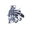
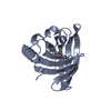
| Structure viewer | Molecule: MolmilJmol/JSmol |
|---|
- Downloads & links
-Download
| PDBx/mmCIF format | 1hqp.cif.gz | 41.3 KB | Display | PDBx/mmCIF format |
|---|---|---|---|---|
| PDB format | pdb1hqp.ent.gz | 28.3 KB | Display | PDB format |
| PDBx/mmJSON format | 1hqp.json.gz | Tree view | PDBx/mmJSON format | |
| Others |  Other downloads Other downloads |
-Validation report
| Arichive directory | https://data.pdbj.org/pub/pdb/validation_reports/hq/1hqpftp://data.pdbj.org/pub/pdb/validation_reports/hq/1hqp | HTTPS FTP |
|---|
-Related structure data
| Related structure data | 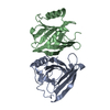 1a3yS S: Starting model for refinement |
|---|---|
| Similar structure data |


-Links
 PDBj
PDBj

- Assembly
| Deposited unit | 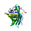
| ||||||||
|---|---|---|---|---|---|---|---|---|---|
| 1 |
| ||||||||
| Unit cell |
| ||||||||
| Details | The asymmetric unit is a monomer. The dimer is generated by the crystallographic two-fold axis |
-Components
| #1: Protein | Mass: 17721.414 Da / Num. of mol.: 1 / Source method: isolated from a natural source / Source: (natural) |
|---|---|
| #2: Chemical | ChemComp-PRZ / 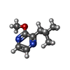  Mass: 166.220 Da / Num. of mol.: 1 / Source method: obtained synthetically / Formula: C9H14N2O Mass: 166.220 Da / Num. of mol.: 1 / Source method: obtained synthetically / Formula: C9H14N2O |
| Has protein modification | Y |
-Experimental details
-Experiment
| Experiment | Method: X-RAY DIFFRACTION / Number of used crystals: 1 |
|---|
- Sample preparation
| Crystal | Density Matthews: 2.19 Å3/Da / Density % sol: 43.81 % | |||||||||||||||||||||||||
|---|---|---|---|---|---|---|---|---|---|---|---|---|---|---|---|---|---|---|---|---|---|---|---|---|---|---|
| Crystal grow | Temperature: 293 K / Method: microdialysis / pH: 7.5 Details: Ammonium sulfate, Tris, ethylene glycol, pH 7.5, MICRODIALYSIS, temperature 293K | |||||||||||||||||||||||||
| Crystal | *PLUS Density % sol: 44 % | |||||||||||||||||||||||||
| Crystal grow | *PLUS Temperature: 20 ℃ | |||||||||||||||||||||||||
| Components of the solutions | *PLUS
|
-Data collection
| Diffraction | Mean temperature: 293 K |
|---|---|
| Diffraction source | Source: ROTATING ANODE / Type: RIGAKU RU200 / Wavelength: 1.5418 Å |
| Detector | Type: XENTRONICS / Detector: AREA DETECTOR / Date: Feb 15, 1992 / Details: graphite monochromator |
| Radiation | Monochromator: Graphite / Protocol: SINGLE WAVELENGTH / Monochromatic (M) / Laue (L): M / Scattering type: x-ray |
| Radiation wavelength | Wavelength: 1.5418 Å / Relative weight: 1 |
| Reflection | Resolution: 2.3→10 Å / Num. all: 14835 / Num. obs: 5662 / % possible obs: 80 % / Observed criterion σ(F): 0 / Observed criterion σ(I): 0 / Redundancy: 2.16 % / Biso Wilson estimate: 37 Å2 / Rmerge(I) obs: 0.046 / Net I/σ(I): 31 |
| Reflection shell | Resolution: 2.3→2.5 Å / Redundancy: 2.2 % / Rmerge(I) obs: 0.107 / Mean I/σ(I) obs: 10.1 / Num. unique all: 1005 / % possible all: 64 |
| Reflection | *PLUS % possible obs: 80 % / Num. measured all: 14835 / Rmerge(I) obs: 0.0466 |
| Reflection shell | *PLUS % possible obs: 64 % / Num. unique obs: 1005 / Rmerge(I) obs: 0.1072 |
- Processing
| Software |
| |||||||||||||||||||||||||
|---|---|---|---|---|---|---|---|---|---|---|---|---|---|---|---|---|---|---|---|---|---|---|---|---|---|---|
| Refinement | Method to determine structure: MOLECULAR REPLACEMENT Starting model: PDB ENTRY 1A3Y Resolution: 2.3→10 Å / σ(F): 0 / σ(I): 0 / Stereochemistry target values: Engh & Huber
| |||||||||||||||||||||||||
| Refinement step | Cycle: LAST / Resolution: 2.3→10 Å
| |||||||||||||||||||||||||
| Refine LS restraints |
| |||||||||||||||||||||||||
| Software | *PLUS Name: TNT / Classification: refinement | |||||||||||||||||||||||||
| Refinement | *PLUS Highest resolution: 2.3 Å / σ(F): 0 | |||||||||||||||||||||||||
| Solvent computation | *PLUS | |||||||||||||||||||||||||
| Displacement parameters | *PLUS | |||||||||||||||||||||||||
| Refine LS restraints | *PLUS
|
