 Movie
Movie Controller


+ Open data

- Basic information
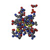
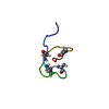
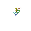
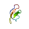
| Entry | Database: PDB / ID: 1hp3 | ||||||
|---|---|---|---|---|---|---|---|
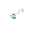
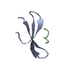
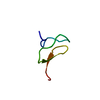
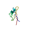
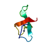
| Title | C-TERMINAL TRUNCATION OF OMEGA-ATRACOTOXIN-HV2A (CT-HV2A) | ||||||
 Components Components | OMEGA-ATRACOTOXIN-HV2A | ||||||
 Keywords Keywords | TOXIN / Cystine knot | ||||||
| Function / homology | Omega-atracotoxin, conserved site-2 / Omega-atracotoxin (ACTX) type 2 family signature. / Magi 5 toxic peptide / Magi 5 toxic peptide family / sodium channel inhibitor activity / calcium channel inhibitor activity / toxin activity / extracellular region / Omega-hexatoxin-Hv2a Function and homology information Function and homology information | ||||||
| Method | SOLUTION NMR / Torsion angle dynamics, dynamical simulated annealing | ||||||
 Authors Authors | Wang, X.-H. / King, G.F. | ||||||
 Citation Citation | Journal: J.Biol.Chem. / Year: 2001 Title: Discovery and structure of a potent and highly specific blocker of insect calcium channels Authors: Wang, X.-H. / Connor, M. / Wilson, D. / Wilson, H.I. / Nicholson, G.M. / Smith, R. / Shaw, D. / Mackay, J.P. / Alewood, P.F. / Christie, M.J. / King, G.F. #1: Journal: Nat.Struct.Biol. / Year: 2000Title: Discovery and characterization of a family of insecticidal neurotoxins with a rare vicinal disulfide bridge. Authors: Wang, X.-H. / Connor, M. / Smith, R. / Maciejewski, M.W. / Howden, M.E.H. / Nicholson, G.M. #2: Journal: Nat.Struct.Biol. / Year: 1997Title: The structure of a novel insecticidal neurotoxin, omega-atracotoxin-HV1, from the venom of an Australian funnel web spider. Authors: Fletcher, J.I. / Smith, R. / O'Donoghue, S.I. / Nilges, M. / Connor, M. / Howden, M.E. / Christie, M.J. / King, G.F. | ||||||
| History |
|
- Structure visualization
| Structure viewer | Molecule: MolmilJmol/JSmol |
|---|
- Downloads & links
-Download
| PDBx/mmCIF format | 1hp3.cif.gz | 179.9 KB | Display | PDBx/mmCIF format |
|---|---|---|---|---|
| PDB format | pdb1hp3.ent.gz | 147.9 KB | Display | PDB format |
| PDBx/mmJSON format | 1hp3.json.gz | Tree view | PDBx/mmJSON format | |
| Others |  Other downloads Other downloads |
-Validation report
| Arichive directory | https://data.pdbj.org/pub/pdb/validation_reports/hp/1hp3ftp://data.pdbj.org/pub/pdb/validation_reports/hp/1hp3 | HTTPS FTP |
|---|
-Related structure data
-Links
 PDBj
PDBj- Assembly
| Deposited unit | 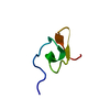
| |||||||||
|---|---|---|---|---|---|---|---|---|---|---|
| 1 |
| |||||||||
| NMR ensembles |
|
-Components
| #1: Protein/peptide | Mass: 3383.948 Da / Num. of mol.: 1 / Fragment: N-TERMINAL (RESIDUES 1-32) / Source method: obtained synthetically Details: This sequence does not occur naturally. The peptide was chemically synthesized using standard t-Boc chemistry, and oxidized/folded in a glutathione redox buffer. References: UniProt: P82852 |
|---|---|
| Has protein modification | Y |
-Experimental details
-Experiment
| Experiment | Method: SOLUTION NMR | ||||||||||||||||
|---|---|---|---|---|---|---|---|---|---|---|---|---|---|---|---|---|---|
| NMR experiment |
| ||||||||||||||||
| NMR details | Text: This structure was determined using standard 2D homonuclear NMR techniques. |
- Sample preparation
| Details |
| |||||||||
|---|---|---|---|---|---|---|---|---|---|---|
| Sample conditions | Ionic strength: 0.005 / pH: 4.71 / Pressure: 1 atm / Temperature: 296 K |
-NMR measurement
| Radiation | Protocol: SINGLE WAVELENGTH / Monochromatic (M) / Laue (L): M |
|---|---|
| Radiation wavelength | Relative weight: 1 |
| NMR spectrometer | Type: Varian INOVA / Manufacturer: Varian / Model: INOVA / Field strength: 600 MHz |
- Processing
| NMR software |
| ||||||||||||||||||||||||
|---|---|---|---|---|---|---|---|---|---|---|---|---|---|---|---|---|---|---|---|---|---|---|---|---|---|
| Refinement | Method: Torsion angle dynamics, dynamical simulated annealing Software ordinal: 1 Details: The structures are based on a total of 345 NOE-derived distance restraints, 21 dihedral-angle restraints, plus 22 restraints defining 11 hydrogen bonds. | ||||||||||||||||||||||||
| NMR representative | Selection criteria: lowest energy | ||||||||||||||||||||||||
| NMR ensemble | Conformer selection criteria: structures with the lowest energy Conformers calculated total number: 100 / Conformers submitted total number: 20 |
 X-PLOR