 Movie
Movie Controller

[English] 日本語
 Yorodumi
- PDB-1hny: The structure of human pancreatic alpha-amylase at 1.8 angstroms ... -
+ Open data

- Basic information
| Entry | Database: PDB / ID: 1hny | |||||||||
|---|---|---|---|---|---|---|---|---|---|---|
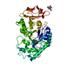
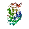
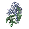
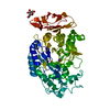
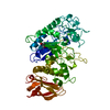
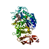
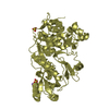
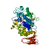
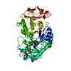
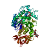
| Title | The structure of human pancreatic alpha-amylase at 1.8 angstroms resolution and comparisons with related enzymes | |||||||||
 Components Components | HUMAN PANCREATIC ALPHA-AMYLASE | |||||||||
 Keywords Keywords | HYDROLASE (O-GLYCOSYL) | |||||||||
| Function / homology |  Function and homology information Function and homology informationpolysaccharide digestion / Digestion of dietary carbohydrate / Developmental Lineage of Pancreatic Acinar Cells / alpha-amylase / alpha-amylase activity / carbohydrate catabolic process / chloride ion binding / carbohydrate metabolic process / calcium ion binding / : ...polysaccharide digestion / Digestion of dietary carbohydrate / Developmental Lineage of Pancreatic Acinar Cells / alpha-amylase / alpha-amylase activity / carbohydrate catabolic process / chloride ion binding / carbohydrate metabolic process / calcium ion binding / : / extracellular exosome / extracellular region Similarity search - Function | |||||||||
| Biological species |  Homo sapiens (human) Homo sapiens (human) | |||||||||
| Method |  X-RAY DIFFRACTION / Resolution: 1.8 Å X-RAY DIFFRACTION / Resolution: 1.8 Å | |||||||||
 Authors Authors | Luo, Y. / Brayer, G.D. | |||||||||
 Citation Citation | Journal: Protein Sci. / Year: 1995 Title: The structure of human pancreatic alpha-amylase at 1.8 A resolution and comparisons with related enzymes. Authors: Brayer, G.D. / Luo, Y. / Withers, S.G. #1: Journal: J.Mol.Biol. / Year: 1993Title: Isolation, Crystallization and Preliminary Diffraction Analyses of Human Pancreatic Alpha-Amylase Authors: Burk, D. / Wang, Y. / Dombroski, D. / Berghuis, A.M. / Evans, S.V. / Luo, Y. / Withers, S.G. / Brayer, G.D. | |||||||||
| History |
|
- Structure visualization
| Structure viewer | Molecule: MolmilJmol/JSmol |
|---|
- Downloads & links
-Download
| PDBx/mmCIF format | 1hny.cif.gz | 119.3 KB | Display | PDBx/mmCIF format |
|---|---|---|---|---|
| PDB format | pdb1hny.ent.gz | 90.7 KB | Display | PDB format |
| PDBx/mmJSON format | 1hny.json.gz | Tree view | PDBx/mmJSON format | |
| Others |  Other downloads Other downloads |
-Validation report
| Arichive directory | https://data.pdbj.org/pub/pdb/validation_reports/hn/1hnyftp://data.pdbj.org/pub/pdb/validation_reports/hn/1hny | HTTPS FTP |
|---|
-Related structure data
| Similar structure data |
|---|
-Links
 PDBj
PDBj

- Assembly
| Deposited unit | 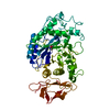
| ||||||||
|---|---|---|---|---|---|---|---|---|---|
| 1 |
| ||||||||
| Unit cell |
| ||||||||
| Atom site foot note | 1: CIS PROLINE - PRO 54 / 2: CIS PROLINE - PRO 130 |
-Components
| #1: Protein | Mass: 55931.305 Da / Num. of mol.: 1 / Source method: isolated from a natural source / Source: (natural) Homo sapiens (human) / Organ: PANCREAS / References: UniProt: P04746, alpha-amylase |
|---|---|
| #2: Chemical | ChemComp-CA /   Mass: 40.078 Da / Num. of mol.: 1 / Source method: obtained synthetically / Formula: Ca Mass: 40.078 Da / Num. of mol.: 1 / Source method: obtained synthetically / Formula: Ca |
| #3: Chemical | ChemComp-CL /   Mass: 35.453 Da / Num. of mol.: 1 / Source method: obtained synthetically / Formula: Cl Mass: 35.453 Da / Num. of mol.: 1 / Source method: obtained synthetically / Formula: Cl |
| #4: Water | ChemComp-HOH /  Mass: 18.015 Da / Num. of mol.: 359 / Source method: isolated from a natural source / Formula: H2O Mass: 18.015 Da / Num. of mol.: 359 / Source method: isolated from a natural source / Formula: H2O |
| Has protein modification | Y |
-Experimental details
-Experiment
| Experiment | Method: X-RAY DIFFRACTION |
|---|
- Sample preparation
| Crystal | Density Matthews: 2.43 Å3/Da / Density % sol: 49.48 % | ||||||||||||||||||||||||||||||
|---|---|---|---|---|---|---|---|---|---|---|---|---|---|---|---|---|---|---|---|---|---|---|---|---|---|---|---|---|---|---|---|
| Crystal | *PLUS Density % sol: 50 % | ||||||||||||||||||||||||||||||
| Crystal grow | *PLUS Temperature: 22 ℃ / pH: 7.5 / Method: vapor diffusion, hanging drop | ||||||||||||||||||||||||||||||
| Components of the solutions | *PLUS
|
-Data collection
| Diffraction source | Wavelength: 1.5418 |
|---|---|
| Detector | Type: RIGAKU / Detector: IMAGE PLATE |
| Radiation | Monochromatic (M) / Laue (L): M / Scattering type: x-ray |
| Radiation wavelength | Wavelength: 1.5418 Å / Relative weight: 1 |
| Reflection | Resolution: 1.8→36.6 Å / Num. obs: 42316 / % possible obs: 82.1 % / Observed criterion σ(I): 2 / Redundancy: 3 % / Rmerge(I) obs: 0.055 |
| Reflection | *PLUS Rmerge(I) obs: 0.055 |
| Reflection shell | *PLUS Highest resolution: 1.8 Å / Lowest resolution: 2 Å / % possible obs: 63.1 % |
- Processing
| Software |
| ||||||||||||||||||||||||||||||||||||||||||||||||||||||||||||||||||||||||||||||||
|---|---|---|---|---|---|---|---|---|---|---|---|---|---|---|---|---|---|---|---|---|---|---|---|---|---|---|---|---|---|---|---|---|---|---|---|---|---|---|---|---|---|---|---|---|---|---|---|---|---|---|---|---|---|---|---|---|---|---|---|---|---|---|---|---|---|---|---|---|---|---|---|---|---|---|---|---|---|---|---|---|---|
| Refinement | Resolution: 1.8→8 Å / σ(F): 2
| ||||||||||||||||||||||||||||||||||||||||||||||||||||||||||||||||||||||||||||||||
| Displacement parameters | Biso mean: 22.71 Å2 | ||||||||||||||||||||||||||||||||||||||||||||||||||||||||||||||||||||||||||||||||
| Refine analyze | Luzzati coordinate error obs: 0.18 Å | ||||||||||||||||||||||||||||||||||||||||||||||||||||||||||||||||||||||||||||||||
| Refinement step | Cycle: LAST / Resolution: 1.8→8 Å
| ||||||||||||||||||||||||||||||||||||||||||||||||||||||||||||||||||||||||||||||||
| Refine LS restraints |
| ||||||||||||||||||||||||||||||||||||||||||||||||||||||||||||||||||||||||||||||||
| Software | *PLUS Name: X-PLOR / Classification: refinement | ||||||||||||||||||||||||||||||||||||||||||||||||||||||||||||||||||||||||||||||||
| Refinement | *PLUS | ||||||||||||||||||||||||||||||||||||||||||||||||||||||||||||||||||||||||||||||||
| Solvent computation | *PLUS | ||||||||||||||||||||||||||||||||||||||||||||||||||||||||||||||||||||||||||||||||
| Displacement parameters | *PLUS | ||||||||||||||||||||||||||||||||||||||||||||||||||||||||||||||||||||||||||||||||
| Refine LS restraints | *PLUS
|
