 Movie
Movie Controller

[English] 日本語
 Yorodumi
- PDB-1hmc: THREE-DIMENSIONAL STRUCTURE OF DIMERIC HUMAN RECOMBINANT MACROPHA... -
+ Open data

- Basic information
| Entry | Database: PDB / ID: 1hmc | ||||||
|---|---|---|---|---|---|---|---|
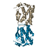
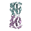
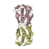
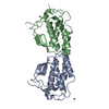
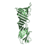
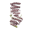
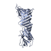
| Title | THREE-DIMENSIONAL STRUCTURE OF DIMERIC HUMAN RECOMBINANT MACROPHAGE COLONY STIMULATING FACTOR | ||||||
 Components Components | HUMAN MACROPHAGE COLONY STIMULATING FACTOR | ||||||
 Keywords Keywords | MACROPHAGE COLONY STIMULATING FACTOR | ||||||
| Function / homology |  Function and homology information Function and homology informationregulation of macrophage derived foam cell differentiation / mammary gland fat development / positive regulation of macrophage colony-stimulating factor signaling pathway / macrophage homeostasis / macrophage colony-stimulating factor receptor binding / monocyte homeostasis / osteoclast proliferation / positive regulation of macrophage migration / positive regulation of odontogenesis of dentin-containing tooth / CSF1-CSF1R complex ...regulation of macrophage derived foam cell differentiation / mammary gland fat development / positive regulation of macrophage colony-stimulating factor signaling pathway / macrophage homeostasis / macrophage colony-stimulating factor receptor binding / monocyte homeostasis / osteoclast proliferation / positive regulation of macrophage migration / positive regulation of odontogenesis of dentin-containing tooth / CSF1-CSF1R complex / monocyte activation / macrophage colony-stimulating factor signaling pathway / developmental process involved in reproduction / positive regulation of microglial cell migration / mammary duct terminal end bud growth / microglial cell proliferation / positive regulation of mononuclear cell proliferation / positive regulation of macrophage differentiation / myeloid leukocyte migration / positive regulation of monocyte differentiation / neutrophil homeostasis / positive regulation of multicellular organism growth / positive regulation of osteoclast differentiation / branching involved in mammary gland duct morphogenesis / positive regulation of macrophage derived foam cell differentiation / positive regulation of Ras protein signal transduction / Other interleukin signaling / positive regulation of cell-matrix adhesion / positive regulation of macrophage chemotaxis / Interleukin-10 signaling / positive regulation of protein metabolic process / macrophage differentiation / regulation of ossification / monocyte differentiation / Transcriptional and post-translational regulation of MITF-M expression and activity / homeostasis of number of cells within a tissue / osteoclast differentiation / cell surface receptor protein tyrosine kinase signaling pathway / response to ischemia / cytokine activity / growth factor activity / Post-translational protein phosphorylation / Regulation of Insulin-like Growth Factor (IGF) transport and uptake by Insulin-like Growth Factor Binding Proteins (IGFBPs) / Signaling by CSF1 (M-CSF) in myeloid cells / Ras protein signal transduction / nuclear body / positive regulation of cell migration / endoplasmic reticulum lumen / inflammatory response / innate immune response / positive regulation of cell population proliferation / positive regulation of gene expression / perinuclear region of cytoplasm / protein homodimerization activity / : / extracellular region / membrane / identical protein binding / plasma membrane Similarity search - Function | ||||||
| Biological species |  Homo sapiens (human) Homo sapiens (human) | ||||||
| Method |  X-RAY DIFFRACTION / Resolution: 2.5 Å X-RAY DIFFRACTION / Resolution: 2.5 Å | ||||||
 Authors Authors | Bohm, A. / Pandit, J. / Jancarik, J. / Halenbeck, R. / Koths, K. / Kim, S.-H. | ||||||
 Citation Citation | Journal: Science / Year: 1992 Title: Three-dimensional structure of dimeric human recombinant macrophage colony-stimulating factor. Authors: Pandit, J. / Bohm, A. / Jancarik, J. / Halenbeck, R. / Koths, K. / Kim, S.H. | ||||||
| History |
|
- Structure visualization
| Structure viewer | Molecule: MolmilJmol/JSmol |
|---|
- Downloads & links
-Download
| PDBx/mmCIF format | 1hmc.cif.gz | 21.2 KB | Display | PDBx/mmCIF format |
|---|---|---|---|---|
| PDB format | pdb1hmc.ent.gz | 10.5 KB | Display | PDB format |
| PDBx/mmJSON format | 1hmc.json.gz | Tree view | PDBx/mmJSON format | |
| Others |  Other downloads Other downloads |
-Validation report
| Arichive directory | https://data.pdbj.org/pub/pdb/validation_reports/hm/1hmcftp://data.pdbj.org/pub/pdb/validation_reports/hm/1hmc | HTTPS FTP |
|---|
-Related structure data
| Similar structure data |
|---|


-Links
 PDBj
PDBj


- Assembly
| Deposited unit | 
| ||||||||
|---|---|---|---|---|---|---|---|---|---|
| 1 |
| ||||||||
| Unit cell |
| ||||||||
| Noncrystallographic symmetry (NCS) | NCS oper: (Code: given Matrix: (-0.925055, -0.377203, -0.043564), Vector: Details | THE TRANSFORMATION PRESENTED ON *MTRIX* RECORDS BELOW WILL YIELD APPROXIMATE COORDINATES FOR CHAIN A WHEN APPLIED TO CHAIN B. | |
-Components
| #1: Protein | Mass: 17292.590 Da / Num. of mol.: 2 Source method: isolated from a genetically manipulated source Source: (gene. exp.) Homo sapiens (human) / References: UniProt: P09603Sequence details | SEQUENCE ADVISORY NOTICE: DIFFERENCE BETWEEN SWISS-PROT AND PDB SEQUENCE. SWISS-PROT ENTRY NAME: : ...SEQUENCE ADVISORY NOTICE: DIFFERENCE | |
|---|
-Experimental details
-Experiment
| Experiment | Method: X-RAY DIFFRACTION |
|---|
- Sample preparation
| Crystal | Density Matthews: 2.47 Å3/Da / Density % sol: 50.11 % | ||||||||||||||||||||||||||||||
|---|---|---|---|---|---|---|---|---|---|---|---|---|---|---|---|---|---|---|---|---|---|---|---|---|---|---|---|---|---|---|---|
| Crystal grow | *PLUS Method: vapor diffusion | ||||||||||||||||||||||||||||||
| Components of the solutions | *PLUS
|
-Data collection
| Radiation | Scattering type: x-ray |
|---|---|
| Radiation wavelength | Relative weight: 1 |
| Reflection | *PLUS Highest resolution: 2 Å / Num. obs: 18842 / Num. measured all: 65871 / Rmerge(I) obs: 0.0701 |
- Processing
| Software |
| ||||||||||||||||||||||||||||||||||||||||||||||||||||||||||||
|---|---|---|---|---|---|---|---|---|---|---|---|---|---|---|---|---|---|---|---|---|---|---|---|---|---|---|---|---|---|---|---|---|---|---|---|---|---|---|---|---|---|---|---|---|---|---|---|---|---|---|---|---|---|---|---|---|---|---|---|---|---|
| Refinement | Resolution: 2.5→8 Å / Rfactor Rwork: 0.197 / Rfactor obs: 0.197 / σ(F): 2 Details: THE SEQUENCE THAT WAS CRYSTALLIZED RAN FROM RESIDUE 4 THROUGH RESIDUE 158. NEITHER OF THE TWO TERMINI COULD BE LOCATED IN THE ELECTRON DENSITY MAPS. THE TWO CRYSTALLOGRAPHICALLY UNIQUE ...Details: THE SEQUENCE THAT WAS CRYSTALLIZED RAN FROM RESIDUE 4 THROUGH RESIDUE 158. NEITHER OF THE TWO TERMINI COULD BE LOCATED IN THE ELECTRON DENSITY MAPS. THE TWO CRYSTALLOGRAPHICALLY UNIQUE MONOMERS HAVE BEEN ASSIGNED CHAIN INDICATORS *A* AND *B*. | ||||||||||||||||||||||||||||||||||||||||||||||||||||||||||||
| Refinement step | Cycle: LAST / Resolution: 2.5→8 Å
| ||||||||||||||||||||||||||||||||||||||||||||||||||||||||||||
| Refine LS restraints |
| ||||||||||||||||||||||||||||||||||||||||||||||||||||||||||||
| Software | *PLUS Name: X-PLOR / Classification: refinement | ||||||||||||||||||||||||||||||||||||||||||||||||||||||||||||
| Refinement | *PLUS Highest resolution: 2.5 Å / Lowest resolution: 8 Å / σ(F): 2 / Rfactor obs: 0.197 | ||||||||||||||||||||||||||||||||||||||||||||||||||||||||||||
| Solvent computation | *PLUS | ||||||||||||||||||||||||||||||||||||||||||||||||||||||||||||
| Displacement parameters | *PLUS | ||||||||||||||||||||||||||||||||||||||||||||||||||||||||||||
| Refine LS restraints | *PLUS Type: x_angle_d / Dev ideal: 3.7 |
