 Movie
Movie Controller

[English] 日本語
 Yorodumi
- PDB-1hlz: CRYSTAL STRUCTURE OF THE ORPHAN NUCLEAR RECEPTOR REV-ERB(ALPHA) D... -
+ Open data

- Basic information
| Entry | Database: PDB / ID: 1hlz | ||||||
|---|---|---|---|---|---|---|---|
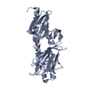
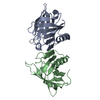
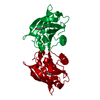
| Title | CRYSTAL STRUCTURE OF THE ORPHAN NUCLEAR RECEPTOR REV-ERB(ALPHA) DNA-BINDING DOMAIN BOUND TO ITS COGNATE RESPONSE ELEMENT | ||||||
 Components Components |
| ||||||
 Keywords Keywords | TRANSCRIPTION/DNA / ORPHAN RECEPTOR / NUCLEAR RECEPTOR / DNA-BINDING / REVERB / REV-ERB / TRANSCRIPTION-DNA COMPLEX | ||||||
| Function / homology |  Function and homology information Function and homology informationregulation of circadian sleep/wake cycle / positive regulation of bile acid biosynthetic process / circadian temperature homeostasis / negative regulation of astrocyte activation / regulation of type B pancreatic cell proliferation / negative regulation of microglial cell activation / negative regulation of toll-like receptor 4 signaling pathway / negative regulation of neuroinflammatory response / response to leptin / regulation of insulin secretion involved in cellular response to glucose stimulus ...regulation of circadian sleep/wake cycle / positive regulation of bile acid biosynthetic process / circadian temperature homeostasis / negative regulation of astrocyte activation / regulation of type B pancreatic cell proliferation / negative regulation of microglial cell activation / negative regulation of toll-like receptor 4 signaling pathway / negative regulation of neuroinflammatory response / response to leptin / regulation of insulin secretion involved in cellular response to glucose stimulus / glycogen biosynthetic process / regulation of fat cell differentiation / Phosphorylated BMAL1:CLOCK (ARNTL:CLOCK) activates expression of core clock genes / negative regulation of cold-induced thermogenesis / nuclear steroid receptor activity / E-box binding / regulation of lipid metabolic process / cellular response to interleukin-1 / intracellular receptor signaling pathway / hormone-mediated signaling pathway / proteasomal protein catabolic process / RORA,B,C and NR1D1 (REV-ERBA) regulate gene expression / Expression of BMAL (ARNTL), CLOCK, and NPAS2 / intracellular glucose homeostasis / cholesterol homeostasis / transcription corepressor binding / negative regulation of canonical NF-kappaB signal transduction / RNA polymerase II transcription regulatory region sequence-specific DNA binding / circadian regulation of gene expression / Heme signaling / PPARA activates gene expression / Transcriptional activation of mitochondrial biogenesis / Nuclear Receptor transcription pathway / regulation of circadian rhythm / DNA-binding transcription repressor activity, RNA polymerase II-specific / protein destabilization / nuclear receptor activity / negative regulation of inflammatory response / cellular response to tumor necrosis factor / sequence-specific double-stranded DNA binding / cellular response to lipopolysaccharide / dendritic spine / DNA-binding transcription factor activity, RNA polymerase II-specific / cell differentiation / transcription cis-regulatory region binding / nuclear body / RNA polymerase II cis-regulatory region sequence-specific DNA binding / negative regulation of DNA-templated transcription / heme binding / dendrite / positive regulation of DNA-templated transcription / chromatin / negative regulation of transcription by RNA polymerase II / positive regulation of transcription by RNA polymerase II / zinc ion binding / nucleoplasm / nucleus / cytoplasm Similarity search - Function | ||||||
| Biological species |  Homo sapiens (human) Homo sapiens (human) | ||||||
| Method |  X-RAY DIFFRACTION / SYNCHROTRON / MOLECULAR REPLACEMENT / Resolution: 2.8 Å X-RAY DIFFRACTION / SYNCHROTRON / MOLECULAR REPLACEMENT / Resolution: 2.8 Å | ||||||
 Authors Authors | Sierk, M.L. / Zhao, Q. / Rastinejad, F. | ||||||
 Citation Citation | Journal: Biochemistry / Year: 2001 Title: DNA Deformability as a Recognition Feature in the RevErb Response Element Authors: Sierk, M.L. / Zhao, Q. / Rastinejad, F. #1: Journal: Mol.Cell / Year: 1998Title: Structural Elements of an Orphan Nuclear Receptor-DNA Complex Authors: Zhao, Q. / Khorasanizadeh, S. / Miyoshi, Y. / Lazar, M. / Rastinejad, F. | ||||||
| History |
|
- Structure visualization
| Structure viewer | Molecule: MolmilJmol/JSmol |
|---|
- Downloads & links
-Download
| PDBx/mmCIF format | 1hlz.cif.gz | 63.3 KB | Display | PDBx/mmCIF format |
|---|---|---|---|---|
| PDB format | pdb1hlz.ent.gz | 42.4 KB | Display | PDB format |
| PDBx/mmJSON format | 1hlz.json.gz | Tree view | PDBx/mmJSON format | |
| Others |  Other downloads Other downloads |
-Validation report
| Arichive directory | https://data.pdbj.org/pub/pdb/validation_reports/hl/1hlzftp://data.pdbj.org/pub/pdb/validation_reports/hl/1hlz | HTTPS FTP |
|---|
-Related structure data
| Related structure data |  1ga5C  1a6yS S: Starting model for refinement C: citing same article ( |
|---|---|
| Similar structure data |


-Links
 PDBj
PDBj


- Assembly
| Deposited unit | 
| ||||||||
|---|---|---|---|---|---|---|---|---|---|
| 1 |
| ||||||||
| Unit cell |
|
-Components
| #1: DNA chain | Mass: 6142.993 Da / Num. of mol.: 1 / Source method: obtained synthetically / Details: Synthesized optimal DR2 target | ||||||
|---|---|---|---|---|---|---|---|
| #2: DNA chain | Mass: 6124.965 Da / Num. of mol.: 1 / Source method: obtained synthetically Details: Synthesized optimal DR2 target complementary strand | ||||||
| #3: Protein | Mass: 10902.009 Da / Num. of mol.: 2 / Fragment: DNA-BINDING DOMAIN PLUS C-TERMINAL EXTENSION Source method: isolated from a genetically manipulated source Source: (gene. exp.) Homo sapiens (human) / Plasmid: PGEX / Species (production host): Escherichia coli / Production host:  #4: Chemical | ChemComp-ZN /   Mass: 65.409 Da / Num. of mol.: 4 / Source method: obtained synthetically / Formula: Zn Mass: 65.409 Da / Num. of mol.: 4 / Source method: obtained synthetically / Formula: Zn#5: Water | ChemComp-HOH / |  Mass: 18.015 Da / Num. of mol.: 9 / Source method: isolated from a natural source / Formula: H2O Mass: 18.015 Da / Num. of mol.: 9 / Source method: isolated from a natural source / Formula: H2OHas protein modification | Y | |
-Experimental details
-Experiment
| Experiment | Method: X-RAY DIFFRACTION / Number of used crystals: 1 |
|---|
- Sample preparation
| Crystal | Density Matthews: 2.16 Å3/Da / Density % sol: 42.93 % | |||||||||||||||||||||||||||||||||||||||||||||||||
|---|---|---|---|---|---|---|---|---|---|---|---|---|---|---|---|---|---|---|---|---|---|---|---|---|---|---|---|---|---|---|---|---|---|---|---|---|---|---|---|---|---|---|---|---|---|---|---|---|---|---|
| Crystal grow | Temperature: 290 K / Method: vapor diffusion, hanging drop / pH: 7.5 Details: 25% PEG8000, 5 mM MgCl2, 400 mM NaCl, Tris buffer, pH 7.5, VAPOR DIFFUSION, HANGING DROP, temperature 290K | |||||||||||||||||||||||||||||||||||||||||||||||||
| Components of the solutions |
| |||||||||||||||||||||||||||||||||||||||||||||||||
| Crystal grow | *PLUS Temperature: 17 ℃ / Details: Zhao, Q., (1998) Mol. Cell, 1, 849. | |||||||||||||||||||||||||||||||||||||||||||||||||
| Components of the solutions | *PLUS
|
-Data collection
| Diffraction | Mean temperature: 100 K |
|---|---|
| Diffraction source | Source: SYNCHROTRON / Site: NSLS  / Beamline: X4A / Wavelength: 0.92016 Å / Beamline: X4A / Wavelength: 0.92016 Å |
| Detector | Type: FUJI / Detector: IMAGE PLATE / Date: Apr 5, 1997 Details: Double crystal monochromator with sagittally focussed second crystal. Two spherical mirrors. |
| Radiation | Monochromator: Double crystal monochromator with sagittally focussed second crystal Protocol: MAD / Monochromatic (M) / Laue (L): M / Scattering type: x-ray |
| Radiation wavelength | Wavelength: 0.92016 Å / Relative weight: 1 |
| Reflection | Resolution: 2.7→18.62 Å / Num. all: 7671 / Num. obs: 7671 / % possible obs: 97 % / Observed criterion σ(F): 0 / Observed criterion σ(I): 0 / Redundancy: 3.1 % / Biso Wilson estimate: 58.3 Å2 / Rmerge(I) obs: 0.09 / Net I/σ(I): 9 |
| Reflection shell | Resolution: 2.7→2.8 Å / Redundancy: 3.1 % / Rmerge(I) obs: 0.44 / Mean I/σ(I) obs: 2.1 / Num. unique all: 7691 / % possible all: 92.7 |
| Reflection | *PLUS Num. obs: 6854 / % possible obs: 96.6 % / Rmerge(I) obs: 0.078 |
| Reflection shell | *PLUS % possible obs: 93.3 % / Num. unique obs: 732 / Rmerge(I) obs: 0.258 / Mean I/σ(I) obs: 3.18 |
- Processing
| Software |
| |||||||||||||||||||||||||
|---|---|---|---|---|---|---|---|---|---|---|---|---|---|---|---|---|---|---|---|---|---|---|---|---|---|---|
| Refinement | Method to determine structure: MOLECULAR REPLACEMENT Starting model: PDB Entry 1A6Y, residues 132-198 from chain A & B, plus DNA Resolution: 2.8→18.6 Å / Isotropic thermal model: anisotropic / Cross valid method: THROUGHOUT / σ(F): 0 / σ(I): 0 / Stereochemistry target values: Engh & Huber Details: Conjugate-gradient minimization against a maximum-likelihood structure factor target. NCS restraints between upstream and downstream monomers and DNA half-sites used throughout refinement. ...Details: Conjugate-gradient minimization against a maximum-likelihood structure factor target. NCS restraints between upstream and downstream monomers and DNA half-sites used throughout refinement. Harmonic restraints used throughout refinement.
| |||||||||||||||||||||||||
| Displacement parameters | Biso mean: 60.81 Å2
| |||||||||||||||||||||||||
| Refine analyze |
| |||||||||||||||||||||||||
| Refinement step | Cycle: LAST / Resolution: 2.8→18.6 Å
| |||||||||||||||||||||||||
| Refine LS restraints |
| |||||||||||||||||||||||||
| LS refinement shell | Resolution: 2.8→2.9 Å
| |||||||||||||||||||||||||
| Software | *PLUS Name: CNS / Version: 0.9 / Classification: refinement | |||||||||||||||||||||||||
| Refinement | *PLUS Highest resolution: 2.8 Å / Lowest resolution: 18.6 Å / σ(F): 0 | |||||||||||||||||||||||||
| Solvent computation | *PLUS | |||||||||||||||||||||||||
| Displacement parameters | *PLUS | |||||||||||||||||||||||||
| Refine LS restraints | *PLUS
| |||||||||||||||||||||||||
| LS refinement shell | *PLUS Highest resolution: 2.7 Å / Lowest resolution: 2.8 Å |
