 Movie
Movie Controller


+ Open data

- Basic information
| Entry | Database: PDB / ID: 1hkw | ||||||
|---|---|---|---|---|---|---|---|
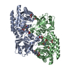
| Title | MYCOBACTERIUM DIAMINOPIMELATE DICARBOXYLASE (LysA) | ||||||
 Components Components | DIAMINOPIMELATE DECARBOXYLASE | ||||||
 Keywords Keywords | LYASE / DECARBOXYLASE / DIAMINOPIMELATE / DAPDC / PLP / LYSINE PATHWAY / MYCOBACTERIUM TUBERCULOSIS / LYSINE SYNTHETIC PATHWAY / PSI / PROTEIN STRUCTURE INITIATIVE / TB STRUCTURAL GENOMICS CONSORTIUM / TB / TBSGC | ||||||
| Function / homology |  Function and homology information Function and homology informationdiaminopimelate decarboxylase / diaminopimelate decarboxylase activity / : / peptidoglycan-based cell wall / pyridoxal phosphate binding Similarity search - Function | ||||||
| Biological species |   MYCOBACTERIUM TUBERCULOSIS (bacteria) MYCOBACTERIUM TUBERCULOSIS (bacteria) | ||||||
| Method |  X-RAY DIFFRACTION / SYNCHROTRON / MAD / Resolution: 2.8 Å X-RAY DIFFRACTION / SYNCHROTRON / MAD / Resolution: 2.8 Å | ||||||
 Authors Authors | Gokulan, K. / Rupp, B. / Pavelka Jr, M.S. / Jacobs Jr, W.R. / Sacchettini, J.C. / TB Structural Genomics Consortium (TBSGC) | ||||||
 Citation Citation | Journal: J.Biol.Chem. / Year: 2003 Title: Crystal Structure of Mycobacterium Tuberculosis Diaminopimelate Decarboxylase, an Essential Enzyme in Bacterial Lysine Biosynthesis Authors: Gokulan, K. / Rupp, B. / Pavelka, M. / Jacobs, W. / Sacchettini, J.C. | ||||||
| History |
| ||||||
| Remark 700 | SHEET THE SHEET STRUCTURE OF THIS MOLECULE IS BIFURCATED. IN ORDER TO REPRESENT THIS FEATURE IN ... SHEET THE SHEET STRUCTURE OF THIS MOLECULE IS BIFURCATED. IN ORDER TO REPRESENT THIS FEATURE IN THE SHEET RECORDS BELOW, TWO SHEETS ARE DEFINED. |
- Structure visualization
| Structure viewer | Molecule: MolmilJmol/JSmol |
|---|
- Downloads & links
-Download
| PDBx/mmCIF format | 1hkw.cif.gz | 178.2 KB | Display | PDBx/mmCIF format |
|---|---|---|---|---|
| PDB format | pdb1hkw.ent.gz | 143.4 KB | Display | PDB format |
| PDBx/mmJSON format | 1hkw.json.gz | Tree view | PDBx/mmJSON format | |
| Others |  Other downloads Other downloads |
-Validation report
| Arichive directory | https://data.pdbj.org/pub/pdb/validation_reports/hk/1hkwftp://data.pdbj.org/pub/pdb/validation_reports/hk/1hkw | HTTPS FTP |
|---|
-Related structure data


-Links
 PDBj
PDBj- Assembly
| Deposited unit | 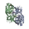
| ||||||||||||||||||
|---|---|---|---|---|---|---|---|---|---|---|---|---|---|---|---|---|---|---|---|
| 1 |
| ||||||||||||||||||
| Unit cell |
| ||||||||||||||||||
| Noncrystallographic symmetry (NCS) | NCS domain:
NCS domain segments: Component-ID: 1 / Ens-ID: 1 / Beg auth comp-ID: GLU / Beg label comp-ID: GLU / End auth comp-ID: GLU / End label comp-ID: GLU / Refine code: 4 / Auth seq-ID: 3 - 445 / Label seq-ID: 3 - 445
NCS oper: (Code: given Matrix: (-0.29392, -0.71332, -0.63623), Vector: |
-Components
| #1: Protein | Mass: 48574.078 Da / Num. of mol.: 2 Source method: isolated from a genetically manipulated source Details: SULFATE ION IN PLP PHOSPHATE BINDING SITE / Source: (gene. exp.) MYCOBACTERIUM TUBERCULOSIS (bacteria) / Strain: H37RV / Description: C-TERM 6-HIS TAG / Production host: References: UniProt: P31848, UniProt: P9WIU7*PLUS, diaminopimelate decarboxylase #2: Chemical |   Mass: 96.063 Da / Num. of mol.: 2 / Source method: obtained synthetically / Formula: SO4 Mass: 96.063 Da / Num. of mol.: 2 / Source method: obtained synthetically / Formula: SO4#3: Water | ChemComp-HOH / |  Mass: 18.015 Da / Num. of mol.: 214 / Source method: isolated from a natural source / Formula: H2O Mass: 18.015 Da / Num. of mol.: 214 / Source method: isolated from a natural source / Formula: H2OCompound details | CONVERTS MESO-2,6-DIAMINOHEPTANEDIOATE TO L-LYSINE AND CO(2). REQUIRES PYRIDOXAL PHOSPHATE AS A ...CONVERTS MESO-2,6-DIAMINOHEP | Has protein modification | Y | Sequence details | RV1293, INITIAL METHIONINE | |
|---|
-Experimental details
-Experiment
| Experiment | Method: X-RAY DIFFRACTION / Number of used crystals: 1 |
|---|
- Sample preparation
| Crystal | Density Matthews: 4.1 Å3/Da / Density % sol: 70 % | ||||||||||||||||||||||||||||||||||||
|---|---|---|---|---|---|---|---|---|---|---|---|---|---|---|---|---|---|---|---|---|---|---|---|---|---|---|---|---|---|---|---|---|---|---|---|---|---|
| Crystal grow | Method: vapor diffusion, hanging drop / pH: 6.5 Details: 4 UL HANGING DROPS (2 UL OF DAPDC 10 MG/ML CONTAINING 5MM OF LYSINE COMBINED WITH 2 UL OF WELL SOLUTION) WERE EQUILIBRATED AGAINST 500 UL OF WELL SOLUTION (24% PEG-MME 5K, 0.1M MES BUFFER ...Details: 4 UL HANGING DROPS (2 UL OF DAPDC 10 MG/ML CONTAINING 5MM OF LYSINE COMBINED WITH 2 UL OF WELL SOLUTION) WERE EQUILIBRATED AGAINST 500 UL OF WELL SOLUTION (24% PEG-MME 5K, 0.1M MES BUFFER PH6.3 AND 60 MM AMMONIUM SULFATE), pH 6.50 | ||||||||||||||||||||||||||||||||||||
| Crystal grow | *PLUS Temperature: 18 ℃ / pH: 6.3 / Method: vapor diffusion, hanging drop | ||||||||||||||||||||||||||||||||||||
| Components of the solutions | *PLUS
|
-Data collection
| Diffraction | Mean temperature: 120 K |
|---|---|
| Diffraction source | Source: SYNCHROTRON / Site: APS  / Beamline: 14-ID-B / Wavelength: 0.9638 / Beamline: 14-ID-B / Wavelength: 0.9638 |
| Detector | Type: ADSC CCD / Detector: CCD / Date: Jun 15, 2002 / Details: MIRROR |
| Radiation | Monochromator: SI / Protocol: SINGLE WAVELENGTH / Monochromatic (M) / Laue (L): M / Scattering type: x-ray |
| Radiation wavelength | Wavelength: 0.9638 Å / Relative weight: 1 |
| Reflection | Resolution: 2.8→65.7 Å / Num. obs: 813664 / % possible obs: 99.9 % / Redundancy: 22.6 % / Rmerge(I) obs: 0.028 / Net I/σ(I): 28.2 |
| Reflection shell | Resolution: 2.8→2.87 Å / Redundancy: 22 % / Rmerge(I) obs: 0.136 / Mean I/σ(I) obs: 7.5 / % possible all: 99.8 |
| Reflection | *PLUS Lowest resolution: 25 Å / Num. obs: 35928 / Num. measured all: 813664 |
| Reflection shell | *PLUS % possible obs: 99.8 % / Num. unique obs: 2637 / Num. measured obs: 58014 |
- Processing
| Software |
| ||||||||||||||||||||||||||||||||||||||||||||||||||||||||||||||||||||||||||||||||||||||||||||||||||||||||||||||||||||||||||||||||||||||||||||||||||||||||||||||||||||||||||||||||||||||
|---|---|---|---|---|---|---|---|---|---|---|---|---|---|---|---|---|---|---|---|---|---|---|---|---|---|---|---|---|---|---|---|---|---|---|---|---|---|---|---|---|---|---|---|---|---|---|---|---|---|---|---|---|---|---|---|---|---|---|---|---|---|---|---|---|---|---|---|---|---|---|---|---|---|---|---|---|---|---|---|---|---|---|---|---|---|---|---|---|---|---|---|---|---|---|---|---|---|---|---|---|---|---|---|---|---|---|---|---|---|---|---|---|---|---|---|---|---|---|---|---|---|---|---|---|---|---|---|---|---|---|---|---|---|---|---|---|---|---|---|---|---|---|---|---|---|---|---|---|---|---|---|---|---|---|---|---|---|---|---|---|---|---|---|---|---|---|---|---|---|---|---|---|---|---|---|---|---|---|---|---|---|---|---|
| Refinement | Method to determine structure: MAD / Resolution: 2.8→25 Å / Cor.coef. Fo:Fc: 0.939 / Cor.coef. Fo:Fc free: 0.899 / SU B: 11.89 / SU ML: 0.223 / Cross valid method: THROUGHOUT / ESU R: 0.496 / ESU R Free: 0.309 / Stereochemistry target values: MAXIMUM LIKELIHOOD Details: HYDROGENS HAVE BEEN ADDED IN THE RIDING POSITIONS.ATOM OXT OF A 447 AND B 446 WERE REMOVED TO INDICATE THAT THE CHAINS CONTINUE BEYOND RESIDUE 447.
| ||||||||||||||||||||||||||||||||||||||||||||||||||||||||||||||||||||||||||||||||||||||||||||||||||||||||||||||||||||||||||||||||||||||||||||||||||||||||||||||||||||||||||||||||||||||
| Solvent computation | Ion probe radii: 0.8 Å / Shrinkage radii: 0.8 Å / VDW probe radii: 1.4 Å / Solvent model: BABINET MODEL WITH MASK | ||||||||||||||||||||||||||||||||||||||||||||||||||||||||||||||||||||||||||||||||||||||||||||||||||||||||||||||||||||||||||||||||||||||||||||||||||||||||||||||||||||||||||||||||||||||
| Displacement parameters | Biso mean: 38.37 Å2
| ||||||||||||||||||||||||||||||||||||||||||||||||||||||||||||||||||||||||||||||||||||||||||||||||||||||||||||||||||||||||||||||||||||||||||||||||||||||||||||||||||||||||||||||||||||||
| Refinement step | Cycle: LAST / Resolution: 2.8→25 Å
| ||||||||||||||||||||||||||||||||||||||||||||||||||||||||||||||||||||||||||||||||||||||||||||||||||||||||||||||||||||||||||||||||||||||||||||||||||||||||||||||||||||||||||||||||||||||
| Refine LS restraints |
|