 Movie
Movie Controller

[English] 日本語
 Yorodumi
- PDB-5x7n: Crystal structure of meso-diaminopimelate decarboxylase (DAPDC) f... -
+ Open data

- Basic information
| Entry | Database: PDB / ID: 5x7n | ||||||
|---|---|---|---|---|---|---|---|
| Title | Crystal structure of meso-diaminopimelate decarboxylase (DAPDC) from Corynebacterium glutamicum | ||||||
 Components Components | Diaminopimelate decarboxylase | ||||||
 Keywords Keywords | LYASE / Decarboxylase | ||||||
| Function / homology |  Function and homology information Function and homology informationdiaminopimelate decarboxylase / diaminopimelate decarboxylase activity / : / pyridoxal phosphate binding Similarity search - Function | ||||||
| Biological species |  Corynebacterium glutamicum (bacteria) Corynebacterium glutamicum (bacteria) | ||||||
| Method |  X-RAY DIFFRACTION / SYNCHROTRON / MOLECULAR REPLACEMENT / Resolution: 1.72 Å X-RAY DIFFRACTION / SYNCHROTRON / MOLECULAR REPLACEMENT / Resolution: 1.72 Å | ||||||
 Authors Authors | Son, H.-F. / Kim, K.-J. | ||||||
 Citation Citation | Journal: Biochem. Biophys. Res. Commun. / Year: 2018 Title: Structural basis for substrate specificity of meso-diaminopimelic acid decarboxylase from Corynebacterium glutamicum. Authors: Son, H.F. / Kim, K.J. | ||||||
| History |
|
- Structure visualization
| Structure viewer | Molecule: MolmilJmol/JSmol |
|---|
- Downloads & links
-Download
| PDBx/mmCIF format | 5x7n.cif.gz | 198.1 KB | Display | PDBx/mmCIF format |
|---|---|---|---|---|
| PDB format | pdb5x7n.ent.gz | 155.1 KB | Display | PDB format |
| PDBx/mmJSON format | 5x7n.json.gz | Tree view | PDBx/mmJSON format | |
| Others |  Other downloads Other downloads |
-Validation report
| Arichive directory | https://data.pdbj.org/pub/pdb/validation_reports/x7/5x7nftp://data.pdbj.org/pub/pdb/validation_reports/x7/5x7n | HTTPS FTP |
|---|
-Related structure data
| Related structure data |  5x7mSC S: Starting model for refinement C: citing same article ( |
|---|---|
| Similar structure data |

-Links
 PDBj
PDBj- Assembly
| Deposited unit | 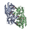
| ||||||||
|---|---|---|---|---|---|---|---|---|---|
| 1 |
| ||||||||
| Unit cell |
|
-Components
| #1: Protein | Mass: 48528.090 Da / Num. of mol.: 2 Source method: isolated from a genetically manipulated source Source: (gene. exp.) Corynebacterium glutamicum (strain ATCC 13032 / DSM 20300 / JCM 1318 / LMG 3730 / NCIMB 10025) (bacteria)Strain: ATCC 13032 / DSM 20300 / JCM 1318 / LMG 3730 / NCIMB 10025 Gene: lysA, Cgl1180, cg1334 / Plasmid: pET30a / Production host: #2: Chemical | ChemComp-GOL /   Mass: 92.094 Da / Num. of mol.: 8 / Source method: obtained synthetically / Formula: C3H8O3 Mass: 92.094 Da / Num. of mol.: 8 / Source method: obtained synthetically / Formula: C3H8O3#3: Chemical |   Mass: 247.142 Da / Num. of mol.: 2 / Source method: obtained synthetically / Formula: C8H10NO6P Mass: 247.142 Da / Num. of mol.: 2 / Source method: obtained synthetically / Formula: C8H10NO6P#4: Chemical |   Type: L-peptide linking / Mass: 147.195 Da / Num. of mol.: 2 / Source method: obtained synthetically / Formula: C6H15N2O2 Type: L-peptide linking / Mass: 147.195 Da / Num. of mol.: 2 / Source method: obtained synthetically / Formula: C6H15N2O2#5: Water | ChemComp-HOH / |  Mass: 18.015 Da / Num. of mol.: 892 / Source method: isolated from a natural source / Formula: H2O Mass: 18.015 Da / Num. of mol.: 892 / Source method: isolated from a natural source / Formula: H2O |
|---|
-Experimental details
-Experiment
| Experiment | Method: X-RAY DIFFRACTION / Number of used crystals: 1 |
|---|
- Sample preparation
| Crystal | Density Matthews: 2.65 Å3/Da / Density % sol: 53.55 % |
|---|---|
| Crystal grow | Temperature: 293 K / Method: vapor diffusion, hanging drop / Details: Sodium citrate tribasic, Sodium cacodylate |
-Data collection
| Diffraction | Mean temperature: 100 K |
|---|---|
| Diffraction source | Source: SYNCHROTRON / Site: PAL/PLS  / Beamline: 7A (6B, 6C1) / Wavelength: 0.97934 Å / Beamline: 7A (6B, 6C1) / Wavelength: 0.97934 Å |
| Detector | Type: ADSC QUANTUM 270 / Detector: CCD / Date: Oct 19, 2014 |
| Radiation | Monochromator: Double Crystal Monochromator / Protocol: SINGLE WAVELENGTH / Monochromatic (M) / Laue (L): M / Scattering type: x-ray |
| Radiation wavelength | Wavelength: 0.97934 Å / Relative weight: 1 |
| Reflection | Resolution: 1.72→95.17 Å / Num. obs: 95643 / % possible obs: 95.2 % / Redundancy: 5.9 % / Net I/σ(I): 40.23 |
| Reflection shell | Resolution: 1.72→1.75 Å |
- Processing
| Software |
| ||||||||||||||||||||||||||||||||||||||||||||||||||||||||||||
|---|---|---|---|---|---|---|---|---|---|---|---|---|---|---|---|---|---|---|---|---|---|---|---|---|---|---|---|---|---|---|---|---|---|---|---|---|---|---|---|---|---|---|---|---|---|---|---|---|---|---|---|---|---|---|---|---|---|---|---|---|---|
| Refinement | Method to determine structure: MOLECULAR REPLACEMENT Starting model: 5X7M Resolution: 1.72→23.461 Å / Cor.coef. Fo:Fc: 0.961 / Cor.coef. Fo:Fc free: 0.943 / SU B: 1.909 / SU ML: 0.062 / Cross valid method: THROUGHOUT / σ(F): 0 / ESU R: 0.104 / ESU R Free: 0.106 / Stereochemistry target values: MAXIMUM LIKELIHOOD Details: HYDROGENS HAVE BEEN ADDED IN THE RIDING POSITIONS U VALUES
| ||||||||||||||||||||||||||||||||||||||||||||||||||||||||||||
| Solvent computation | Ion probe radii: 0.8 Å / Shrinkage radii: 0.8 Å / VDW probe radii: 1.2 Å / Solvent model: MASK | ||||||||||||||||||||||||||||||||||||||||||||||||||||||||||||
| Displacement parameters | Biso max: 86.14 Å2 / Biso mean: 18.718 Å2 / Biso min: 9.44 Å2
| ||||||||||||||||||||||||||||||||||||||||||||||||||||||||||||
| Refinement step | Cycle: final / Resolution: 1.72→23.461 Å
| ||||||||||||||||||||||||||||||||||||||||||||||||||||||||||||
| Refine LS restraints |
| ||||||||||||||||||||||||||||||||||||||||||||||||||||||||||||
| LS refinement shell | Resolution: 1.722→1.767 Å / Rfactor Rfree error: 0 / Total num. of bins used: 20
|
