 Movie
Movie Controller

[English] 日本語
 Yorodumi
- PDB-1hii: COMPARATIVE ANALYSIS OF THE X-RAY STRUCTURES OF HIV-1 AND HIV-2 P... -
+ Open data

- Basic information
| Entry | Database: PDB / ID: 1hii | ||||||
|---|---|---|---|---|---|---|---|
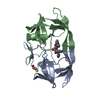
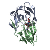
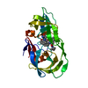
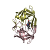
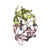
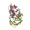
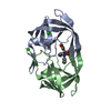
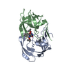
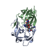
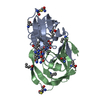
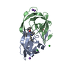
| Title | COMPARATIVE ANALYSIS OF THE X-RAY STRUCTURES OF HIV-1 AND HIV-2 PROTEASES IN COMPLEX WITH CGP 53820, A NOVEL PSEUDOSYMMETRIC INHIBITOR | ||||||
 Components Components | HIV-2 PROTEASE | ||||||
 Keywords Keywords | HYDROLASE (ASPARTIC PROTEINASE) / ASPARTATE PROTEASE / INHIBITED / HIV | ||||||
| Function / homology |  Function and homology information Function and homology informationHIV-2 retropepsin / retroviral ribonuclease H / exoribonuclease H / exoribonuclease H activity / DNA integration / viral genome integration into host DNA / establishment of integrated proviral latency / RNA-directed DNA polymerase / RNA stem-loop binding / viral penetration into host nucleus ...HIV-2 retropepsin / retroviral ribonuclease H / exoribonuclease H / exoribonuclease H activity / DNA integration / viral genome integration into host DNA / establishment of integrated proviral latency / RNA-directed DNA polymerase / RNA stem-loop binding / viral penetration into host nucleus / host multivesicular body / RNA-directed DNA polymerase activity / RNA-DNA hybrid ribonuclease activity / Transferases; Transferring phosphorus-containing groups; Nucleotidyltransferases / host cell / viral nucleocapsid / DNA recombination / DNA-directed DNA polymerase / aspartic-type endopeptidase activity / Hydrolases; Acting on ester bonds / DNA-directed DNA polymerase activity / symbiont-mediated suppression of host gene expression / viral translational frameshifting / symbiont entry into host cell / lipid binding / host cell nucleus / host cell plasma membrane / virion membrane / structural molecule activity / proteolysis / DNA binding / zinc ion binding Similarity search - Function | ||||||
| Biological species |  Human immunodeficiency virus 2 Human immunodeficiency virus 2 | ||||||
| Method |  X-RAY DIFFRACTION / Resolution: 2.3 Å X-RAY DIFFRACTION / Resolution: 2.3 Å | ||||||
 Authors Authors | Priestle, J.P. / Gruetter, M.G. | ||||||
 Citation Citation | Journal: Structure / Year: 1995 Title: Comparative analysis of the X-ray structures of HIV-1 and HIV-2 proteases in complex with CGP 53820, a novel pseudosymmetric inhibitor. Authors: Priestle, J.P. / Fassler, A. / Rosel, J. / Tintelnot-Blomley, M. / Strop, P. / Grutter, M.G. #1: Journal: Bioorg.Med.Chem.Lett. / Year: 1993Title: Novel Pseudosymmetric Inhibitors of HIV-1 Protease Authors: Fassler, A. / Rosel, J. / Tintelnot-Blomley, M. / Alteri, E. / Bold, G. / Lang, M. | ||||||
| History |
|
- Structure visualization
| Structure viewer | Molecule: MolmilJmol/JSmol |
|---|
- Downloads & links
-Download
| PDBx/mmCIF format | 1hii.cif.gz | 55.7 KB | Display | PDBx/mmCIF format |
|---|---|---|---|---|
| PDB format | pdb1hii.ent.gz | 39.6 KB | Display | PDB format |
| PDBx/mmJSON format | 1hii.json.gz | Tree view | PDBx/mmJSON format | |
| Others |  Other downloads Other downloads |
-Validation report
| Arichive directory | https://data.pdbj.org/pub/pdb/validation_reports/hi/1hiiftp://data.pdbj.org/pub/pdb/validation_reports/hi/1hii | HTTPS FTP |
|---|
-Related structure data
-Links
 PDBj
PDBj
- Assembly
| Deposited unit | 
| ||||||||
|---|---|---|---|---|---|---|---|---|---|
| 1 |
| ||||||||
| Unit cell |
| ||||||||
| Noncrystallographic symmetry (NCS) | NCS oper: (Code: given Matrix: (0.26336, -0.90733, 0.32772), Vector: Details | MTRIX THE TRANSFORMATIONS PRESENTED ON MTRIX RECORDS BELOW DESCRIBE NON-CRYSTALLOGRAPHIC RELATIONSHIPS AMONG THE VARIOUS DOMAINS IN THIS ENTRY. APPLYING THE APPROPRIATE MTRIX TRANSFORMATION TO THE RESIDUES LISTED FIRST WILL YIELD APPROXIMATE COORDINATES FOR THE RESIDUES LISTED SECOND. APPLIED TO TRANSFORMED TO MTRIX RESIDUES RESIDUES RMSD M1 A 1 .. A 99 B 1 .. B 99 0.479 | |
-Components
| #1: Protein | Mass: 10728.337 Da / Num. of mol.: 2 Source method: isolated from a genetically manipulated source Source: (gene. exp.) Human immunodeficiency virus 2 / Genus: Lentivirus / Cell line: S2 / Gene: POL / Plasmid: PT7Q10H / Gene (production host): POL / Production host:  References: UniProt: P04584, Hydrolases; Acting on peptide bonds (peptidases); Aspartic endopeptidases #2: Chemical |   Mass: 96.063 Da / Num. of mol.: 2 / Source method: obtained synthetically / Formula: SO4 Mass: 96.063 Da / Num. of mol.: 2 / Source method: obtained synthetically / Formula: SO4#3: Chemical | ChemComp-C20 / |   Mass: 573.767 Da / Num. of mol.: 1 / Source method: obtained synthetically / Formula: C31H51N5O5 Mass: 573.767 Da / Num. of mol.: 1 / Source method: obtained synthetically / Formula: C31H51N5O5#4: Water | ChemComp-HOH / |  Mass: 18.015 Da / Num. of mol.: 194 / Source method: isolated from a natural source / Formula: H2O Mass: 18.015 Da / Num. of mol.: 194 / Source method: isolated from a natural source / Formula: H2OCompound details | COMPND MOLECULE: HIV-2 PROTEASE. ROD ISOLATE. | Nonpolymer details | SOURCE 1 MOLECULE_NAME: CGP 53820. PSEUDOSYMMETRIC TRANSITION-STATE ANALOG. SOURCE 2 MOLECULE_NAME: ...SOURCE 1 MOLECULE_NAME: CGP 53820. PSEUDOSYMM | |
|---|
-Experimental details
-Experiment
| Experiment | Method: X-RAY DIFFRACTION |
|---|
- Sample preparation
| Crystal | Density Matthews: 2.48 Å3/Da / Density % sol: 50.5 % | ||||||||||||||||||||||||||||||
|---|---|---|---|---|---|---|---|---|---|---|---|---|---|---|---|---|---|---|---|---|---|---|---|---|---|---|---|---|---|---|---|
| Crystal | *PLUS Density % sol: 50 % | ||||||||||||||||||||||||||||||
| Crystal grow | *PLUS Method: other | ||||||||||||||||||||||||||||||
| Components of the solutions | *PLUS
|
-Data collection
| Diffraction source | Wavelength: 1.5418 |
|---|---|
| Detector | Type: ENRAF-NONIUS FAST / Detector: DIFFRACTOMETER / Date: Feb 1, 1993 |
| Radiation | Scattering type: x-ray |
| Radiation wavelength | Wavelength: 1.5418 Å / Relative weight: 1 |
| Reflection | Num. obs: 9517 / % possible obs: 94.3 % / Observed criterion σ(I): 0 / Redundancy: 4.13 % / Rmerge(I) obs: 0.051 |
| Reflection | *PLUS Highest resolution: 2.3 Å / Num. measured all: 39279 / Rmerge(I) obs: 0.051 |
| Reflection shell | *PLUS Highest resolution: 2.3 Å / Lowest resolution: 2.38 Å / Rmerge(I) obs: 0.19 |
- Processing
| Software |
| ||||||||||||||||||||||||||||||
|---|---|---|---|---|---|---|---|---|---|---|---|---|---|---|---|---|---|---|---|---|---|---|---|---|---|---|---|---|---|---|---|
| Refinement | Resolution: 2.3→6 Å / σ(F): 0 /
| ||||||||||||||||||||||||||||||
| Displacement parameters | Biso mean: 22.8 Å2 | ||||||||||||||||||||||||||||||
| Refinement step | Cycle: LAST / Resolution: 2.3→6 Å
| ||||||||||||||||||||||||||||||
| Refine LS restraints |
| ||||||||||||||||||||||||||||||
| Software | *PLUS Name: TNT / Classification: refinement | ||||||||||||||||||||||||||||||
| Refine LS restraints | *PLUS
|
