 Movie
Movie Controller


+ Open data

- Basic information
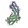
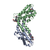
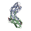
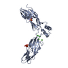
| Entry | Database: PDB / ID: 1h6g | ||||||
|---|---|---|---|---|---|---|---|
| Title | alpha-catenin M-domain | ||||||
 Components Components | ALPHA-1 CATENIN | ||||||
 Keywords Keywords | CELL ADHESION / ALPHA-CATENIN / ADHESION MODULATION / CYTOSKELETON | ||||||
| Function / homology |  Function and homology information Function and homology informationnegative regulation of integrin-mediated signaling pathway / CDH11 homotypic and heterotypic interactions / Regulation of CDH19 Expression and Function / Regulation of CDH11 function / epithelial cell-cell adhesion / zonula adherens / gamma-catenin binding / gap junction assembly / cellular response to indole-3-methanol / vinculin binding ...negative regulation of integrin-mediated signaling pathway / CDH11 homotypic and heterotypic interactions / Regulation of CDH19 Expression and Function / Regulation of CDH11 function / epithelial cell-cell adhesion / zonula adherens / gamma-catenin binding / gap junction assembly / cellular response to indole-3-methanol / vinculin binding / flotillin complex / apical junction assembly / catenin complex / positive regulation of extrinsic apoptotic signaling pathway in absence of ligand / negative regulation of cell motility / positive regulation of smoothened signaling pathway / Regulation of CDH1 Function / Adherens junctions interactions / axon regeneration / negative regulation of neuroblast proliferation / negative regulation of protein localization to nucleus / Myogenesis / smoothened signaling pathway / odontogenesis of dentin-containing tooth / establishment or maintenance of cell polarity / neuroblast proliferation / intercalated disc / sperm head-tail coupling apparatus / negative regulation of extrinsic apoptotic signaling pathway in absence of ligand / ovarian follicle development / RHO GTPases activate IQGAPs / extrinsic apoptotic signaling pathway in absence of ligand / acrosomal vesicle / VEGFR2 mediated vascular permeability / integrin-mediated signaling pathway / adherens junction / cell-cell adhesion / response to estrogen / beta-catenin binding / Degradation of CDH1 / male gonad development / cell-cell junction / actin filament binding / cell junction / intracellular protein localization / cell migration / lamellipodium / actin cytoskeleton / sperm principal piece / sperm midpiece / cell adhesion / cadherin binding / focal adhesion / structural molecule activity / Golgi apparatus / RNA binding / identical protein binding / nucleus / plasma membrane / cytosol Similarity search - Function | ||||||
| Biological species |  HOMO SAPIENS (human) HOMO SAPIENS (human) | ||||||
| Method |  X-RAY DIFFRACTION / SYNCHROTRON / MAD / Resolution: 2.2 Å X-RAY DIFFRACTION / SYNCHROTRON / MAD / Resolution: 2.2 Å | ||||||
 Authors Authors | Yang, J. / Dokurno, P. / Tonks, N.K. / Barford, D. | ||||||
 Citation Citation | Journal: Embo J. / Year: 2001 Title: Crystal Structure of the M-Fragment of Alpha-Catenin: Implications for Modulation of Cell Adhesion. Authors: Yang, J. / Dokurno, P. / Tonks, N.K. / Barford, D. | ||||||
| History |
|
- Structure visualization
| Structure viewer | Molecule: MolmilJmol/JSmol |
|---|
- Downloads & links
-Download
| PDBx/mmCIF format | 1h6g.cif.gz | 116.1 KB | Display | PDBx/mmCIF format |
|---|---|---|---|---|
| PDB format | pdb1h6g.ent.gz | 90.4 KB | Display | PDB format |
| PDBx/mmJSON format | 1h6g.json.gz | Tree view | PDBx/mmJSON format | |
| Others |  Other downloads Other downloads |
-Validation report
| Arichive directory | https://data.pdbj.org/pub/pdb/validation_reports/h6/1h6gftp://data.pdbj.org/pub/pdb/validation_reports/h6/1h6g | HTTPS FTP |
|---|
-Related structure data
| Similar structure data |
|---|


-Links
 PDBj
PDBj


- Assembly
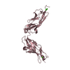
| Deposited unit | 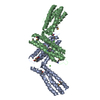
| ||||||||
|---|---|---|---|---|---|---|---|---|---|
| 1 | 
| ||||||||
| 2 | 
| ||||||||
| Unit cell |
|
-Components
| #1: Protein | Mass: 28880.719 Da / Num. of mol.: 2 / Fragment: M-FRAGMENT, RESIDUES 377-632 Source method: isolated from a genetically manipulated source Source: (gene. exp.) HOMO SAPIENS (human) / Plasmid: PET28M / Production host:  #2: Chemical | ChemComp-MPD / ( |   Mass: 118.174 Da / Num. of mol.: 1 / Source method: obtained synthetically / Formula: C6H14O2 / Comment: precipitant*YM Mass: 118.174 Da / Num. of mol.: 1 / Source method: obtained synthetically / Formula: C6H14O2 / Comment: precipitant*YM#3: Chemical |   Mass: 40.078 Da / Num. of mol.: 2 / Source method: obtained synthetically / Formula: Ca Mass: 40.078 Da / Num. of mol.: 2 / Source method: obtained synthetically / Formula: Ca#4: Chemical | ChemComp-CL / |   Mass: 35.453 Da / Num. of mol.: 1 / Source method: obtained synthetically / Formula: Cl Mass: 35.453 Da / Num. of mol.: 1 / Source method: obtained synthetically / Formula: Cl#5: Water | ChemComp-HOH / |  Mass: 18.015 Da / Num. of mol.: 393 / Source method: isolated from a natural source / Formula: H2O Mass: 18.015 Da / Num. of mol.: 393 / Source method: isolated from a natural source / Formula: H2OHas protein modification | Y | |
|---|
-Experimental details
-Experiment
| Experiment | Method: X-RAY DIFFRACTION / Number of used crystals: 1 |
|---|
- Sample preparation
| Crystal | Density Matthews: 2.43 Å3/Da / Density % sol: 49 % | ||||||||||||||||||||||||
|---|---|---|---|---|---|---|---|---|---|---|---|---|---|---|---|---|---|---|---|---|---|---|---|---|---|
| Crystal grow | pH: 4.6 Details: 30 % (V/V) MPD, 0.1 M SODIUM ACETETE BUFFER, PH 4.6, 20 MM CALCIUM CHLORIDE | ||||||||||||||||||||||||
| Crystal grow | *PLUS Temperature: 20 ℃ / Method: vapor diffusion, hanging drop / Details: used microseeding | ||||||||||||||||||||||||
| Components of the solutions | *PLUS
|
-Data collection
| Diffraction | Mean temperature: 100 K |
|---|---|
| Diffraction source | Source: SYNCHROTRON / Site: SRS  / Beamline: PX14.1 / Wavelength: 1.488 / Beamline: PX14.1 / Wavelength: 1.488 |
| Detector | Type: ADSC IMAGE PLATE / Detector: IMAGE PLATE / Date: Sep 8, 2000 |
| Radiation | Protocol: SINGLE WAVELENGTH / Monochromatic (M) / Laue (L): M / Scattering type: x-ray |
| Radiation wavelength | Wavelength: 1.488 Å / Relative weight: 1 |
| Reflection | Resolution: 2.2→30 Å / Num. obs: 27803 / % possible obs: 99.1 % / Redundancy: 6.2 % / Biso Wilson estimate: 28.6 Å2 / Rmerge(I) obs: 0.049 / Net I/σ(I): 13.3 |
| Reflection shell | Resolution: 2.2→2.25 Å / Rmerge(I) obs: 0.205 / % possible all: 98.4 |
| Reflection | *PLUS Num. measured all: 171946 |
| Reflection shell | *PLUS % possible obs: 98.4 % |
- Processing
| Software |
| ||||||||||||||||||||||||||||||||||||||||||||||||||||||||||||||||||||||||||||||||
|---|---|---|---|---|---|---|---|---|---|---|---|---|---|---|---|---|---|---|---|---|---|---|---|---|---|---|---|---|---|---|---|---|---|---|---|---|---|---|---|---|---|---|---|---|---|---|---|---|---|---|---|---|---|---|---|---|---|---|---|---|---|---|---|---|---|---|---|---|---|---|---|---|---|---|---|---|---|---|---|---|---|
| Refinement | Method to determine structure: MAD / Resolution: 2.2→24.74 Å / Rfactor Rfree error: 0.007 / Data cutoff high absF: 11930144.31 / Isotropic thermal model: RESTRAINED / Cross valid method: THROUGHOUT / σ(F): 0 / Details: BULK SOLVENT MODEL USED
| ||||||||||||||||||||||||||||||||||||||||||||||||||||||||||||||||||||||||||||||||
| Solvent computation | Solvent model: FLAT MODEL / Bsol: 87.3308 Å2 / ksol: 0.330463 e/Å3 | ||||||||||||||||||||||||||||||||||||||||||||||||||||||||||||||||||||||||||||||||
| Displacement parameters | Biso mean: 44.6 Å2
| ||||||||||||||||||||||||||||||||||||||||||||||||||||||||||||||||||||||||||||||||
| Refine analyze |
| ||||||||||||||||||||||||||||||||||||||||||||||||||||||||||||||||||||||||||||||||
| Refinement step | Cycle: LAST / Resolution: 2.2→24.74 Å
| ||||||||||||||||||||||||||||||||||||||||||||||||||||||||||||||||||||||||||||||||
| Refine LS restraints |
| ||||||||||||||||||||||||||||||||||||||||||||||||||||||||||||||||||||||||||||||||
| LS refinement shell | Resolution: 2.2→2.34 Å / Rfactor Rfree error: 0.019 / Total num. of bins used: 6
| ||||||||||||||||||||||||||||||||||||||||||||||||||||||||||||||||||||||||||||||||
| Xplor file |
| ||||||||||||||||||||||||||||||||||||||||||||||||||||||||||||||||||||||||||||||||
| Refine LS restraints | *PLUS
| ||||||||||||||||||||||||||||||||||||||||||||||||||||||||||||||||||||||||||||||||
| LS refinement shell | *PLUS Rfactor Rfree: 0.29 / Rfactor Rwork: 0.21 |
