 Movie
Movie Controller

[English] 日本語
 Yorodumi
- PDB-1h5v: Thiopentasaccharide complex of the endoglucanase Cel5A from Bacil... -
+ Open data

- Basic information
| Entry | Database: PDB / ID: 1h5v | |||||||||
|---|---|---|---|---|---|---|---|---|---|---|
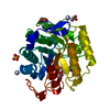
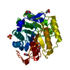
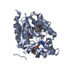
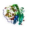
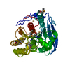
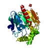
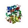
| Title | Thiopentasaccharide complex of the endoglucanase Cel5A from Bacillus agaradharens at 1.1 A resolution in the tetragonal crystal form | |||||||||
 Components Components | ENDOGLUCANASE 5A | |||||||||
 Keywords Keywords | HYDROLASE / CELLULASE / ENDOGLUCANASE / THIOOLIGOSACCHARIDE | |||||||||
| Function / homology |  Function and homology information Function and homology informationcellulase / cellulase activity / cellulose catabolic process / carbohydrate binding / extracellular region Similarity search - Function | |||||||||
| Biological species |  BACILLUS AGARADHAERENS (bacteria) BACILLUS AGARADHAERENS (bacteria) | |||||||||
| Method |  X-RAY DIFFRACTION / SYNCHROTRON / MOLECULAR REPLACEMENT / Resolution: 1.1 Å X-RAY DIFFRACTION / SYNCHROTRON / MOLECULAR REPLACEMENT / Resolution: 1.1 Å | |||||||||
 Authors Authors | Varrot, A. / Sulzenbacher, G. / Schulein, M. / Driguez, H. / Davies, G.J. | |||||||||
 Citation Citation | Journal: Acta Crystallogr.,Sect.D / Year: 2001 Title: Atomic Resolution Structure of Endoglucanase Cel5A in Complex with Methyl 4,4II,4III,4Iv-Tetrathio-Alpha-Cellopentoside Highlights the Alternative Binding Modes Targeted by Substrate Mimics Authors: Varrot, A. / Schulein, M. / Fruchard, S. / Driguez, H. / Davies, G.J. | |||||||||
| History |
| |||||||||
| Remark 700 | SHEET DETERMINATION METHOD: DSSP THE SHEETS PRESENTED AS "AB" IN EACH CHAIN ON SHEET RECORDS BELOW ... SHEET DETERMINATION METHOD: DSSP THE SHEETS PRESENTED AS "AB" IN EACH CHAIN ON SHEET RECORDS BELOW IS ACTUALLY AN 8-STRANDED BARREL THIS IS REPRESENTED BY A 9-STRANDED SHEET IN WHICH THE FIRST AND LAST STRANDS ARE IDENTICAL. |
- Structure visualization
| Structure viewer | Molecule: MolmilJmol/JSmol |
|---|
- Downloads & links
-Download
| PDBx/mmCIF format | 1h5v.cif.gz | 164.4 KB | Display | PDBx/mmCIF format |
|---|---|---|---|---|
| PDB format | pdb1h5v.ent.gz | 127.5 KB | Display | PDB format |
| PDBx/mmJSON format | 1h5v.json.gz | Tree view | PDBx/mmJSON format | |
| Others |  Other downloads Other downloads |
-Validation report
| Arichive directory | https://data.pdbj.org/pub/pdb/validation_reports/h5/1h5vftp://data.pdbj.org/pub/pdb/validation_reports/h5/1h5v | HTTPS FTP |
|---|
-Related structure data
| Related structure data |  1qhzS S: Starting model for refinement |
|---|---|
| Similar structure data |


-Links
 PDBj
PDBj- Assembly
| Deposited unit | 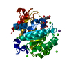
| ||||||||
|---|---|---|---|---|---|---|---|---|---|
| 1 |
| ||||||||
| Unit cell |
| ||||||||
| Components on special symmetry positions |
|
-Components
| #1: Protein | Mass: 34118.168 Da / Num. of mol.: 1 / Fragment: CATALYTIC CORE DOMAIN RESIDUES 27-331 Source method: isolated from a genetically manipulated source Source: (gene. exp.) BACILLUS AGARADHAERENS (bacteria) / Plasmid: THERMAMYL-AMYLASE PROMOT / Production host: | ||||||
|---|---|---|---|---|---|---|---|
| #2: Polysaccharide | alpha-D-glucopyranose-(1-4)-1,4-dithio-beta-D-glucopyranose-(1-4)-4-thio-beta-D-glucopyranose-(1-4)- ...alpha-D-glucopyranose-(1-4)-1,4-dithio-beta-D-glucopyranose-(1-4)-4-thio-beta-D-glucopyranose-(1-4)-4-thio-beta-D-glucopyranose-(1-4)-methyl 4-thio-alpha-D-glucopyranoside Type: oligosaccharide / Mass: 923.072 Da / Num. of mol.: 1 Source method: isolated from a genetically manipulated source | ||||||
| #3: Chemical | ChemComp-CA /   Mass: 40.078 Da / Num. of mol.: 10 / Source method: obtained synthetically / Formula: Ca Mass: 40.078 Da / Num. of mol.: 10 / Source method: obtained synthetically / Formula: Ca#4: Chemical |   Mass: 22.990 Da / Num. of mol.: 2 / Source method: obtained synthetically / Formula: Na Mass: 22.990 Da / Num. of mol.: 2 / Source method: obtained synthetically / Formula: Na#5: Water | ChemComp-HOH / |  Mass: 18.015 Da / Num. of mol.: 537 / Source method: isolated from a natural source / Formula: H2O Mass: 18.015 Da / Num. of mol.: 537 / Source method: isolated from a natural source / Formula: H2OSequence details | THE FIRST 26 RESIDUES IN THE DATABASE CORRESPOND TO THE PROSEQUENCE. OUR NUMBERING BEGINS AT THE ...THE FIRST 26 RESIDUES IN THE DATABASE CORRESPOND | |
-Experimental details
-Experiment
| Experiment | Method: X-RAY DIFFRACTION / Number of used crystals: 1 |
|---|
- Sample preparation
| Crystal | Density Matthews: 2.73 Å3/Da / Density % sol: 54.6 % | ||||||||||||||||||||||||||||
|---|---|---|---|---|---|---|---|---|---|---|---|---|---|---|---|---|---|---|---|---|---|---|---|---|---|---|---|---|---|
| Crystal grow | pH: 7 Details: PROTEIN (20MGML-1) WAS CRYSTALLISED FROM 28% PEG400 AS PRECIPITANT, 100MM HEPES AT PH 7.0 AS BUFFER AND 200 MM CACL2. THE PROTEIN WAS INCUBATED WITH THE 1MM OF SUBSTRATE FOR AN HOUR PRIOR TO COCRYSTALLISATIOM | ||||||||||||||||||||||||||||
| Crystal grow | *PLUS pH: 7 / Method: unknown | ||||||||||||||||||||||||||||
| Components of the solutions | *PLUS
|
-Data collection
| Diffraction | Mean temperature: 100 K |
|---|---|
| Diffraction source | Source: SYNCHROTRON / Site: ESRF  / Beamline: ID14-2 / Wavelength: 0.933 / Beamline: ID14-2 / Wavelength: 0.933 |
| Detector | Detector: CCD / Date: Mar 15, 2000 / Details: TORROIDAL MIRROR |
| Radiation | Monochromator: DIAMOND(111)GE(220) / Protocol: SINGLE WAVELENGTH / Monochromatic (M) / Laue (L): M / Scattering type: x-ray |
| Radiation wavelength | Wavelength: 0.933 Å / Relative weight: 1 |
| Reflection | Resolution: 1.1→20 Å / Num. obs: 155682 / % possible obs: 99.5 % / Redundancy: 3.6 % / Rmerge(I) obs: 0.055 / Net I/σ(I): 20.6 |
| Reflection shell | Resolution: 1.1→1.14 Å / Redundancy: 3.2 % / Rmerge(I) obs: 0.288 / Mean I/σ(I) obs: 4.2 / % possible all: 98.7 |
| Reflection | *PLUS Highest resolution: 1.1 Å / Lowest resolution: 20 Å / Redundancy: 3.6 % |
| Reflection shell | *PLUS % possible obs: 98.7 % / Redundancy: 3.2 % / Mean I/σ(I) obs: 4.2 |
- Processing
| Software |
| ||||||||||||||||||||
|---|---|---|---|---|---|---|---|---|---|---|---|---|---|---|---|---|---|---|---|---|---|
| Refinement | Method to determine structure: MOLECULAR REPLACEMENT Starting model: PDB ENTRY 1QHZ Resolution: 1.1→20 Å / SU B: 0.72227 / SU ML: 0.01778 / Cross valid method: THROUGHOUT / σ(F): 0 / ESU R Free: 0.03223 / Details: THE FIRST THREE RESIDUES DUE TO DISORD
| ||||||||||||||||||||
| Refinement step | Cycle: LAST / Resolution: 1.1→20 Å
| ||||||||||||||||||||
| Refinement | *PLUS Lowest resolution: 20 Å / Rfactor obs: 0.12 / Rfactor Rfree: 0.138 / Rfactor Rwork: 0.12 | ||||||||||||||||||||
| Solvent computation | *PLUS | ||||||||||||||||||||
| Displacement parameters | *PLUS | ||||||||||||||||||||
| Refine LS restraints | *PLUS
|
