 Movie
Movie Controller


+ Open data

- Basic information
| Entry | Database: PDB / ID: 1h5q | ||||||
|---|---|---|---|---|---|---|---|
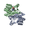
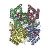
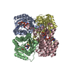
| Title | Mannitol dehydrogenase from Agaricus bisporus | ||||||
 Components Components | NADP-DEPENDENT MANNITOL DEHYDROGENASE | ||||||
 Keywords Keywords | OXIDOREDUCTASE / MANNITOL METABOLISM | ||||||
| Function / homology |  Function and homology information Function and homology informationmannitol 2-dehydrogenase (NADP+) / mannitol metabolic process / mannitol 2-dehydrogenase (NADP+) activity / oxidoreductase activity, acting on NAD(P)H, oxygen as acceptor / NADP binding / protein homotetramerization Similarity search - Function | ||||||
| Biological species |  AGARICUS BISPORUS (cultivated mushroom) AGARICUS BISPORUS (cultivated mushroom) | ||||||
| Method |  X-RAY DIFFRACTION / SYNCHROTRON / MOLECULAR REPLACEMENT / Resolution: 1.5 Å X-RAY DIFFRACTION / SYNCHROTRON / MOLECULAR REPLACEMENT / Resolution: 1.5 Å | ||||||
 Authors Authors | Horer, S. / Stoop, J. / Mooibroek, H. / Baumann, U. / Sassoon, J. | ||||||
 Citation Citation | Journal: J.Biol.Chem. / Year: 2001 Title: The Crystallographic Structure of the Mannitol 2-Dehydrogenase Nadp+ Binary Complex from Agaricus Bisporus Authors: Horer, S. / Stoop, J. / Mooibroek, H. / Baumann, U. / Sassoon, J. #1: Journal: Acta Crystallogr.,Sect.D / Year: 2001 Title: Crystallization and Preliminary Crystallographic Analysis of Mannitol Dehydrogenase (Mtdh) from the Common Mushroom Agaricus Bisporus Authors: Sassoon, J. / Horer, S. / Stoop, J. / Mooibroek, H. / Baumann, U. | ||||||
| History |
|
- Structure visualization
| Structure viewer | Molecule: MolmilJmol/JSmol |
|---|
- Downloads & links
-Download
| PDBx/mmCIF format | 1h5q.cif.gz | 1.2 MB | Display | PDBx/mmCIF format |
|---|---|---|---|---|
| PDB format | pdb1h5q.ent.gz | 1 MB | Display | PDB format |
| PDBx/mmJSON format | 1h5q.json.gz | Tree view | PDBx/mmJSON format | |
| Others |  Other downloads Other downloads |
-Validation report
| Arichive directory | https://data.pdbj.org/pub/pdb/validation_reports/h5/1h5qftp://data.pdbj.org/pub/pdb/validation_reports/h5/1h5q | HTTPS FTP |
|---|
-Related structure data
| Related structure data |  1cydS S: Starting model for refinement |
|---|---|
| Similar structure data |


-Links
 PDBj
PDBj

- Assembly
| Deposited unit | 
| ||||||||||||||||||||||||||||||||||||||||||||||||
|---|---|---|---|---|---|---|---|---|---|---|---|---|---|---|---|---|---|---|---|---|---|---|---|---|---|---|---|---|---|---|---|---|---|---|---|---|---|---|---|---|---|---|---|---|---|---|---|---|---|
| 1 | 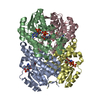
| ||||||||||||||||||||||||||||||||||||||||||||||||
| 2 | 
| ||||||||||||||||||||||||||||||||||||||||||||||||
| 3 | 
| ||||||||||||||||||||||||||||||||||||||||||||||||
| Unit cell |
| ||||||||||||||||||||||||||||||||||||||||||||||||
| Noncrystallographic symmetry (NCS) | NCS oper:
|
-Components
| #1: Protein | Mass: 28237.947 Da / Num. of mol.: 12 Source method: isolated from a genetically manipulated source Source: (gene. exp.) AGARICUS BISPORUS (cultivated mushroom)Plasmid: PET28 / Production host:  References: UniProt: O93868, mannitol 2-dehydrogenase (NADP+) #2: Chemical | ChemComp-NAP /   Mass: 743.405 Da / Num. of mol.: 12 / Source method: obtained synthetically / Formula: C21H28N7O17P3 Mass: 743.405 Da / Num. of mol.: 12 / Source method: obtained synthetically / Formula: C21H28N7O17P3#3: Chemical | ChemComp-NI /   Mass: 58.693 Da / Num. of mol.: 6 / Source method: obtained synthetically / Formula: Ni Mass: 58.693 Da / Num. of mol.: 6 / Source method: obtained synthetically / Formula: Ni#4: Water | ChemComp-HOH / |  Mass: 18.015 Da / Num. of mol.: 3191 / Source method: isolated from a natural source / Formula: H2O Mass: 18.015 Da / Num. of mol.: 3191 / Source method: isolated from a natural source / Formula: H2O |
|---|
-Experimental details
-Experiment
| Experiment | Method: X-RAY DIFFRACTION / Number of used crystals: 1 |
|---|
- Sample preparation
| Crystal | Density Matthews: 2.4 Å3/Da / Density % sol: 47 % | ||||||||||||||||||||||||||||||||||||
|---|---|---|---|---|---|---|---|---|---|---|---|---|---|---|---|---|---|---|---|---|---|---|---|---|---|---|---|---|---|---|---|---|---|---|---|---|---|
| Crystal grow | pH: 7.5 / Details: 90 MM TRIS-HCL PH 7.5, 18% PEG4000, 9% ISOPROPANOL | ||||||||||||||||||||||||||||||||||||
| Crystal grow | *PLUS Temperature: 20 ℃ / Method: vapor diffusion, sitting drop / pH: 7.5 | ||||||||||||||||||||||||||||||||||||
| Components of the solutions | *PLUS
|
-Data collection
| Diffraction | Mean temperature: 120 K |
|---|---|
| Diffraction source | Source: SYNCHROTRON / Site: ESRF  / Beamline: BM1A / Wavelength: 0.8 / Beamline: BM1A / Wavelength: 0.8 |
| Detector | Type: MAR scanner 345 mm plate / Detector: IMAGE PLATE / Date: Jul 15, 2000 |
| Radiation | Protocol: SINGLE WAVELENGTH / Monochromatic (M) / Laue (L): M / Scattering type: x-ray |
| Radiation wavelength | Wavelength: 0.8 Å / Relative weight: 1 |
| Reflection | Resolution: 1.5→40 Å / Num. obs: 508943 / % possible obs: 98.4 % / Redundancy: 2.7 % / Rmerge(I) obs: 0.063 / Net I/σ(I): 11.8 |
| Reflection shell | Resolution: 1.5→1.58 Å / Redundancy: 2.4 % / Rmerge(I) obs: 0.295 / Mean I/σ(I) obs: 3.1 / % possible all: 97 |
| Reflection | *PLUS Lowest resolution: 40 Å |
| Reflection shell | *PLUS % possible obs: 97 % / Num. unique obs: 173816 |
- Processing
| Software |
| ||||||||||||||||||||
|---|---|---|---|---|---|---|---|---|---|---|---|---|---|---|---|---|---|---|---|---|---|
| Refinement | Method to determine structure: MOLECULAR REPLACEMENT Starting model: 1CYD Resolution: 1.5→20 Å / SU B: 1.45 / SU ML: 0.05 / Cross valid method: THROUGHOUT / ESU R Free: 0.07 / Details: HYDROGENS HAVE BEEN ADDED IN THE RIDING POSITIONS
| ||||||||||||||||||||
| Refinement step | Cycle: LAST / Resolution: 1.5→20 Å
| ||||||||||||||||||||
| Software | *PLUS Name: REFMAC / Classification: refinement | ||||||||||||||||||||
| Refine LS restraints | *PLUS
|
