 Movie
Movie Controller

[English] 日本語
 Yorodumi
- PDB-1h56: Structural and biochemical characterization of a new magnesium io... -
+ Open data

- Basic information
| Entry | Database: PDB / ID: 1h56 | ||||||
|---|---|---|---|---|---|---|---|
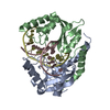
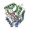
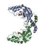
| Title | Structural and biochemical characterization of a new magnesium ion binding site near Tyr94 in the restriction endonuclease PvuII | ||||||
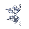
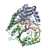
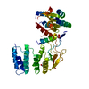
 Components Components | TYPE II RESTRICTION ENZYME PVUII | ||||||
 Keywords Keywords | ENDONUCLEASE / TYPE II RESTRICTION ENDONUCLEASE / HYDROLASE / NUCLEASE | ||||||
| Function / homology |  Function and homology information Function and homology informationtype II site-specific deoxyribonuclease / type II site-specific deoxyribonuclease activity / DNA restriction-modification system / DNA binding / metal ion binding Similarity search - Function | ||||||
| Biological species |  PROTEUS VULGARIS (bacteria) PROTEUS VULGARIS (bacteria) | ||||||
| Method |  X-RAY DIFFRACTION / SYNCHROTRON / FOURIER SYNTHESIS / Resolution: 3 Å X-RAY DIFFRACTION / SYNCHROTRON / FOURIER SYNTHESIS / Resolution: 3 Å | ||||||
 Authors Authors | Spyrida, A. / Matzen, C. / Lanio, T. / Jeltsch, A. / Simoncsits, A. / Athanasiadis, A. / Scheuring-Vanamee, E. / Kokkinidis, M. / Pingoud, A. | ||||||
 Citation Citation | Journal: J.Mol.Biol. / Year: 2003 Title: Structural and Biochemical Characterization of a New Mg(2+) Binding Site Near Tyr94 in the Restriction Endonuclease PvuII. Authors: Spyridaki, A. / Matzen, C. / Lanio, T. / Jeltsch, A. / Simoncsits, A. / Athanasiadis, A. / Scheuring-Vanamee, E. / Kokkinidis, M. / Pingoud, A. #1: Journal: Nat.Struct.Biol. / Year: 1994Title: Crystal Structure of PvuII Endonuclease Reveals Extensive Structural Homologies to EcoRV Authors: Athanasiadis, A. / Vlassi, M. / Kotsifaki, D. / Tucker, P.A. / Wilson, K.S. / Kokkinidis, M. | ||||||
| History |
|
- Structure visualization
| Structure viewer | Molecule: MolmilJmol/JSmol |
|---|
- Downloads & links
-Download
| PDBx/mmCIF format | 1h56.cif.gz | 77.9 KB | Display | PDBx/mmCIF format |
|---|---|---|---|---|
| PDB format | pdb1h56.ent.gz | 58.7 KB | Display | PDB format |
| PDBx/mmJSON format | 1h56.json.gz | Tree view | PDBx/mmJSON format | |
| Others |  Other downloads Other downloads |
-Validation report
| Arichive directory | https://data.pdbj.org/pub/pdb/validation_reports/h5/1h56ftp://data.pdbj.org/pub/pdb/validation_reports/h5/1h56 | HTTPS FTP |
|---|
-Related structure data
| Related structure data | 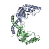 1pvuS S: Starting model for refinement |
|---|---|
| Similar structure data |


-Links
 PDBj
PDBj


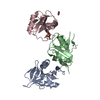
- Assembly
| Deposited unit | 
| ||||||||
|---|---|---|---|---|---|---|---|---|---|
| 1 |
| ||||||||
| Unit cell |
|
-Components
| #1: Protein | Mass: 18239.799 Da / Num. of mol.: 2 Source method: isolated from a genetically manipulated source Source: (gene. exp.) PROTEUS VULGARIS (bacteria) / Plasmid: PPVU1 / Production host: References: UniProt: P23657, type II site-specific deoxyribonuclease #2: Chemical |   Mass: 24.305 Da / Num. of mol.: 2 / Source method: obtained synthetically / Formula: Mg Mass: 24.305 Da / Num. of mol.: 2 / Source method: obtained synthetically / Formula: Mg#3: Water | ChemComp-HOH / |  Mass: 18.015 Da / Num. of mol.: 68 / Source method: isolated from a natural source / Formula: H2O Mass: 18.015 Da / Num. of mol.: 68 / Source method: isolated from a natural source / Formula: H2O |
|---|
-Experimental details
-Experiment
| Experiment | Method: X-RAY DIFFRACTION / Number of used crystals: 1 |
|---|
- Sample preparation
| Crystal | Density Matthews: 2.83 Å3/Da / Density % sol: 56.61 % |
|---|---|
| Crystal grow | pH: 5 / Details: pH 5.00 |
| Crystal grow | *PLUS Method: other / Details: Athanasiadis, A., (1991) J. Mol. Biol., 222, 451. |
-Data collection
| Diffraction | Mean temperature: 293 K |
|---|---|
| Diffraction source | Source: SYNCHROTRON / Site: EMBL/DESY, HAMBURG  / Beamline: X11 / Wavelength: 0.92 / Beamline: X11 / Wavelength: 0.92 |
| Detector | Type: MARRESEARCH / Detector: IMAGE PLATE / Date: Oct 15, 1997 |
| Radiation | Protocol: SINGLE WAVELENGTH / Monochromatic (M) / Laue (L): M / Scattering type: x-ray |
| Radiation wavelength | Wavelength: 0.92 Å / Relative weight: 1 |
| Reflection | Resolution: 3→8 Å / Num. obs: 8104 / % possible obs: 92.3 % / Redundancy: 1.8 % / Rmerge(I) obs: 0.13 |
| Reflection | *PLUS Highest resolution: 3 Å / Lowest resolution: 8 Å / Rmerge(I) obs: 0.13 |
- Processing
| Software |
| ||||||||||||||||||||||||||||||||||||||||||||||||||||||||||||||||||||||||||||||||
|---|---|---|---|---|---|---|---|---|---|---|---|---|---|---|---|---|---|---|---|---|---|---|---|---|---|---|---|---|---|---|---|---|---|---|---|---|---|---|---|---|---|---|---|---|---|---|---|---|---|---|---|---|---|---|---|---|---|---|---|---|---|---|---|---|---|---|---|---|---|---|---|---|---|---|---|---|---|---|---|---|---|
| Refinement | Method to determine structure: FOURIER SYNTHESIS Starting model: PDB ENTRY 1PVU Resolution: 3→8 Å / Rfactor Rfree error: 0.009 / Cross valid method: THROUGHOUT / σ(F): 0
| ||||||||||||||||||||||||||||||||||||||||||||||||||||||||||||||||||||||||||||||||
| Refine analyze |
| ||||||||||||||||||||||||||||||||||||||||||||||||||||||||||||||||||||||||||||||||
| Refinement step | Cycle: LAST / Resolution: 3→8 Å
| ||||||||||||||||||||||||||||||||||||||||||||||||||||||||||||||||||||||||||||||||
| Refine LS restraints |
| ||||||||||||||||||||||||||||||||||||||||||||||||||||||||||||||||||||||||||||||||
| LS refinement shell | Resolution: 3→3.18 Å / Rfactor Rfree error: 0.029 / Total num. of bins used: 6
| ||||||||||||||||||||||||||||||||||||||||||||||||||||||||||||||||||||||||||||||||
| Xplor file |
| ||||||||||||||||||||||||||||||||||||||||||||||||||||||||||||||||||||||||||||||||
| Refinement | *PLUS Highest resolution: 3 Å / Lowest resolution: 8 Å / % reflection Rfree: 10 % / Rfactor Rfree: 0.264 / Rfactor Rwork: 0.153 | ||||||||||||||||||||||||||||||||||||||||||||||||||||||||||||||||||||||||||||||||
| Solvent computation | *PLUS | ||||||||||||||||||||||||||||||||||||||||||||||||||||||||||||||||||||||||||||||||
| Displacement parameters | *PLUS | ||||||||||||||||||||||||||||||||||||||||||||||||||||||||||||||||||||||||||||||||
| Refine LS restraints | *PLUS
|
