 Movie
Movie Controller

[English] 日本語
 Yorodumi
- PDB-1f0o: PVUII ENDONUCLEASE/COGNATE DNA COMPLEX (GLUTARALDEHYDE-CROSSLINKE... -
+ Open data

- Basic information
| Entry | Database: PDB / ID: 1f0o | ||||||
|---|---|---|---|---|---|---|---|
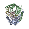
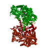
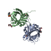
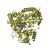
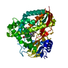
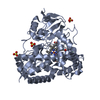
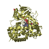
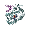
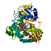
| Title | PVUII ENDONUCLEASE/COGNATE DNA COMPLEX (GLUTARALDEHYDE-CROSSLINKED CRYSTAL) AT PH 7.5 WITH TWO CALCIUM IONS AT EACH ACTIVE SITE | ||||||
 Components Components |
| ||||||
 Keywords Keywords | hydrolase/DNA / PROTEIN-DNA COMPLEX / ENDONUCLEASE TYPE II / RESTRICTION ENZYME / Catalytic Metal visualization / hydrolase-DNA COMPLEX | ||||||
| Function / homology |  Function and homology information Function and homology informationtype II site-specific deoxyribonuclease / type II site-specific deoxyribonuclease activity / DNA restriction-modification system / DNA binding / metal ion binding Similarity search - Function | ||||||
| Biological species |  Proteus vulgaris (bacteria) Proteus vulgaris (bacteria) | ||||||
| Method |  X-RAY DIFFRACTION / Resolution: 2.5 Å X-RAY DIFFRACTION / Resolution: 2.5 Å | ||||||
 Authors Authors | Horton, J.R. / Cheng, X. | ||||||
 Citation Citation | Journal: J.Mol.Biol. / Year: 2000 Title: PvuII endonuclease contains two calcium ions in active sites. Authors: Horton, J.R. / Cheng, X. #1: Journal: J.Mol.Biol. / Year: 1998Title: Asp34 of PvuII Endonuclease is directly involved in DNA minor groove recognition and indirectly involved in catalysis Authors: Horton, J.R. / Nastri, H.G. / Riggs, P.D. / Cheng, X. #2: Journal: Biol.Chem. / Year: 1998Title: How is modification of the DNA substrate recognized by the PvuII restriction endonuclease? Authors: Horton, J.R. / Bonventre, J. / Cheng, X. | ||||||
| History |
|
- Structure visualization
| Structure viewer | Molecule: MolmilJmol/JSmol |
|---|
- Downloads & links
-Download
| PDBx/mmCIF format | 1f0o.cif.gz | 94.5 KB | Display | PDBx/mmCIF format |
|---|---|---|---|---|
| PDB format | pdb1f0o.ent.gz | 68.6 KB | Display | PDB format |
| PDBx/mmJSON format | 1f0o.json.gz | Tree view | PDBx/mmJSON format | |
| Others |  Other downloads Other downloads |
-Validation report
| Arichive directory | https://data.pdbj.org/pub/pdb/validation_reports/f0/1f0oftp://data.pdbj.org/pub/pdb/validation_reports/f0/1f0o | HTTPS FTP |
|---|
-Related structure data


-Links
 PDBj
PDBj


- Assembly
| Deposited unit | 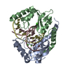
| ||||||||||
|---|---|---|---|---|---|---|---|---|---|---|---|
| 1 |
| ||||||||||
| Unit cell |
| ||||||||||
| Details | The biological assembly is a dimer consisting of protein chain A and chain B and double strained DNA consisting of chains C and D with two CA+2 at each monomeric active site. |
-Components
| #1: DNA chain | Mass: 3967.585 Da / Num. of mol.: 2 / Source method: obtained synthetically Details: SELF-ANNEALING OLIGONUCLEOTIDE CONTAINING COGNATE SIX BASE PAIR SEQUENCE #2: Protein | Mass: 18370.992 Da / Num. of mol.: 2 Source method: isolated from a genetically manipulated source Source: (gene. exp.) Proteus vulgaris (bacteria) / Plasmid: PPR594 / Production host: References: UniProt: P23657, type II site-specific deoxyribonuclease #3: Chemical | ChemComp-CA /   Mass: 40.078 Da / Num. of mol.: 4 / Source method: obtained synthetically / Formula: Ca Mass: 40.078 Da / Num. of mol.: 4 / Source method: obtained synthetically / Formula: Ca#4: Water | ChemComp-HOH / |  Mass: 18.015 Da / Num. of mol.: 190 / Source method: isolated from a natural source / Formula: H2O Mass: 18.015 Da / Num. of mol.: 190 / Source method: isolated from a natural source / Formula: H2O |
|---|
-Experimental details
-Experiment
| Experiment | Method: X-RAY DIFFRACTION / Number of used crystals: 1 |
|---|
- Sample preparation
| Crystal | Density Matthews: 2.03 Å3/Da / Density % sol: 39.55 % | ||||||||||||||||||||||||||||||||||||||||||||||||
|---|---|---|---|---|---|---|---|---|---|---|---|---|---|---|---|---|---|---|---|---|---|---|---|---|---|---|---|---|---|---|---|---|---|---|---|---|---|---|---|---|---|---|---|---|---|---|---|---|---|
| Crystal grow | Temperature: 289 K / Method: vapor diffusion, hanging drop / pH: 4.5 Details: PEG 4000, SODIUM ACETATE, CaCL2, pH 4.5, VAPOR DIFFUSION, HANGING DROP, temperature 289K | ||||||||||||||||||||||||||||||||||||||||||||||||
| Components of the solutions |
| ||||||||||||||||||||||||||||||||||||||||||||||||
| Crystal grow | *PLUS Temperature: 16 ℃Details: Balendiran, K., (1994) Proteins Struct.Funct.Genet., 19, 77. | ||||||||||||||||||||||||||||||||||||||||||||||||
| Components of the solutions | *PLUS
|
-Data collection
| Diffraction | Mean temperature: 95 K |
|---|---|
| Diffraction source | Source: ROTATING ANODE / Type: RIGAKU RU300 / Wavelength: 1.5418 |
| Detector | Type: RIGAKU RAXIS IV / Detector: IMAGE PLATE / Date: Feb 14, 1999 |
| Radiation | Protocol: SINGLE WAVELENGTH / Monochromatic (M) / Laue (L): M / Scattering type: x-ray |
| Radiation wavelength | Wavelength: 1.5418 Å / Relative weight: 1 |
| Reflection | Resolution: 2.5→30 Å / Num. all: 13150 / Num. obs: 13150 / % possible obs: 98.9 % / Observed criterion σ(I): -3 / Redundancy: 4.7 % / Biso Wilson estimate: 23.7 Å2 / Rmerge(I) obs: 0.057 / Net I/σ(I): 20.1 |
| Reflection shell | Resolution: 2.5→2.54 Å / Redundancy: 3.1 % / Rmerge(I) obs: 0.332 / % possible all: 96.5 |
| Reflection | *PLUS Num. measured all: 61937 |
- Processing
| Software |
| |||||||||||||||||||||||||
|---|---|---|---|---|---|---|---|---|---|---|---|---|---|---|---|---|---|---|---|---|---|---|---|---|---|---|
| Refinement | Resolution: 2.5→30 Å / Cross valid method: THROUGHOUT / σ(F): 0 / σ(I): 0 / Stereochemistry target values: Engh & Huber
| |||||||||||||||||||||||||
| Refinement step | Cycle: LAST / Resolution: 2.5→30 Å
| |||||||||||||||||||||||||
| Refine LS restraints |
| |||||||||||||||||||||||||
| Software | *PLUS Name: X-PLOR / Version: 3.851 / Classification: refinement | |||||||||||||||||||||||||
| Refinement | *PLUS Highest resolution: 2.5 Å / Lowest resolution: 30 Å / σ(F): 0 / % reflection Rfree: 10 % / Rfactor obs: 0.205 | |||||||||||||||||||||||||
| Solvent computation | *PLUS | |||||||||||||||||||||||||
| Displacement parameters | *PLUS | |||||||||||||||||||||||||
| Refine LS restraints | *PLUS Type: x_angle_deg / Dev ideal: 1.3 |
