 Movie
Movie Controller

[English] 日本語
 Yorodumi
- PDB-1h2d: Ebola virus matrix protein VP40 N-terminal domain in complex with... -
+ Open data

- Basic information
| Entry | Database: PDB / ID: 1h2d | ||||||
|---|---|---|---|---|---|---|---|
| Title | Ebola virus matrix protein VP40 N-terminal domain in complex with RNA (Low-resolution VP40[31-212] variant). | ||||||
 Components Components |
| ||||||
 Keywords Keywords | VIRUS/VIRAL PROTEIN / FILOVIRUS / EBOLA VIRUS / MATRIX PROTEIN VP40 / ASSEMBLY / BUDDING / VIRUS-VIRAL PROTEIN complex | ||||||
| Function / homology |  Function and homology information Function and homology informationintracellular transport of virus / symbiont-mediated suppression of host RNAi-mediated antiviral immune response / host cell endomembrane system / viral budding / symbiont-mediated perturbation of host cell cycle progression / host cell late endosome membrane / viral budding via host ESCRT complex / host cell membrane / viral budding from plasma membrane / host cell ...intracellular transport of virus / symbiont-mediated suppression of host RNAi-mediated antiviral immune response / host cell endomembrane system / viral budding / symbiont-mediated perturbation of host cell cycle progression / host cell late endosome membrane / viral budding via host ESCRT complex / host cell membrane / viral budding from plasma membrane / host cell / structural constituent of virion / membrane raft / ribonucleoprotein complex / host cell plasma membrane / virion membrane / RNA binding / extracellular region / identical protein binding Similarity search - Function | ||||||
| Biological species |   EBOLA VIRUS EBOLA VIRUS | ||||||
| Method |  X-RAY DIFFRACTION / SYNCHROTRON / MOLECULAR REPLACEMENT / Resolution: 2.6 Å X-RAY DIFFRACTION / SYNCHROTRON / MOLECULAR REPLACEMENT / Resolution: 2.6 Å | ||||||
 Authors Authors | Gomis-Ruth, F.X. / Dessen, A. / Bracher, A. / Klenk, H.D. / Weissenhorn, W. | ||||||
 Citation Citation | Journal: Structure / Year: 2003 Title: The Matrix Protein Vp40 from Ebola Virus Octamerizes Into Pore-Like Structures with Specific RNA Binding Properties Authors: Gomis-Ruth, F.X. / Dessen, A. / Timmins, J. / Bracher, A. / Kolesnikowa, L. / Becker, S. / Klenk, H.D. / Weissenhorn, W. #1: Journal: Embo J. / Year: 2000Title: Crystal Structure of the Matrix Protein Vp40 from Ebola Virus Authors: Dessen, A. / Volchkov, V. / Dolnik, O. / Klenk, H.D. / Weissenhorn, W. | ||||||
| History |
| ||||||
| Remark 700 | SHEET THE SHEET STRUCTURE OF THIS MOLECULE IS BIFURCATED. IN ORDER TO REPRESENT THIS FEATURE IN ... SHEET THE SHEET STRUCTURE OF THIS MOLECULE IS BIFURCATED. IN ORDER TO REPRESENT THIS FEATURE IN THE SHEET RECORDS BELOW, TWO SHEETS ARE DEFINED. |
- Structure visualization
| Structure viewer | Molecule: MolmilJmol/JSmol |
|---|
- Downloads & links
-Download
| PDBx/mmCIF format | 1h2d.cif.gz | 69.9 KB | Display | PDBx/mmCIF format |
|---|---|---|---|---|
| PDB format | pdb1h2d.ent.gz | 50.1 KB | Display | PDB format |
| PDBx/mmJSON format | 1h2d.json.gz | Tree view | PDBx/mmJSON format | |
| Others |  Other downloads Other downloads |
-Validation report
| Arichive directory | https://data.pdbj.org/pub/pdb/validation_reports/h2/1h2dftp://data.pdbj.org/pub/pdb/validation_reports/h2/1h2d | HTTPS FTP |
|---|
-Related structure data
| Related structure data |  1h2cSC S: Starting model for refinement C: citing same article ( |
|---|---|
| Similar structure data |


-Links
 PDBj
PDBj
- Assembly
| Deposited unit | 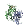
| ||||||||
|---|---|---|---|---|---|---|---|---|---|
| 1 | 
| ||||||||
| Unit cell |
| ||||||||
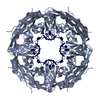
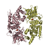
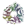
| Details | THE BIOLOGICALLY RELEVANT UNIT COMPRISES AN OCTAMEROF THE PROTEIN WHICH IS IN COMPLEX WITH RNA IN THISENTRY MAKING AN HEXADECAMER. SEE REMARK 400 BELOW |
-Components
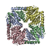
| #1: Protein | Mass: 19655.395 Da / Num. of mol.: 2 / Fragment: N-TERMINAL DOMAIN, RESIDUES 31-212 Source method: isolated from a genetically manipulated source Details: VP40[31-212] VARIANT / Source: (gene. exp.) EBOLA VIRUS / Strain: ZAIRE MAYINGA / Production host: #2: RNA chain | Mass: 935.620 Da / Num. of mol.: 2 / Source method: isolated from a natural source Details: MRNA STOP-CODON SEQUENCE, BIOCHEMICAL SYNTHESIS BY THE EXPRESSION HOST AND UPTAKE BY THE PROTEIN DURING OVEREXPRESSION Source: (natural) #3: Chemical |   Mass: 35.453 Da / Num. of mol.: 2 / Source method: obtained synthetically / Formula: Cl Mass: 35.453 Da / Num. of mol.: 2 / Source method: obtained synthetically / Formula: Cl#4: Water | ChemComp-HOH / |  Mass: 18.015 Da / Num. of mol.: 109 / Source method: isolated from a natural source / Formula: H2O Mass: 18.015 Da / Num. of mol.: 109 / Source method: isolated from a natural source / Formula: H2OCompound details | THE BIOLOGICALLY RELEVANT OLIGOMER IS A HOMOOCTAMER AS CREATED BY THE CRYSTALLOGRAPHIC 222 SYMMETRY ...THE BIOLOGICAL | |
|---|
-Experimental details
-Experiment
| Experiment | Method: X-RAY DIFFRACTION |
|---|
- Sample preparation
| Crystal | Density Matthews: 2.7 Å3/Da / Density % sol: 55 % / Description: PROTEIN DIMER USED AS SEARCH MODEL. | |||||||||||||||||||||||||||||||||||
|---|---|---|---|---|---|---|---|---|---|---|---|---|---|---|---|---|---|---|---|---|---|---|---|---|---|---|---|---|---|---|---|---|---|---|---|---|
| Crystal grow | Method: vapor diffusion, hanging drop / pH: 4.6 Details: 1 UL OF PROTEIN (13 MG/ML) AND 1 UL OF WELL SOLUTION (100 MM NA-ACETATE PH4.6, 35 % MPD, 4 % GLYCEROL).HANGING DROP., pH 4.60 | |||||||||||||||||||||||||||||||||||
| Crystal grow | *PLUS pH: 7.5 / Method: vapor diffusion | |||||||||||||||||||||||||||||||||||
| Components of the solutions | *PLUS
|
-Data collection
| Diffraction | Mean temperature: 100 K |
|---|---|
| Diffraction source | Source: SYNCHROTRON / Site: ESRF  / Beamline: ID14-4 / Wavelength: 0.979 / Beamline: ID14-4 / Wavelength: 0.979 |
| Radiation | Protocol: SINGLE WAVELENGTH / Monochromatic (M) / Laue (L): M / Scattering type: x-ray |
| Radiation wavelength | Wavelength: 0.979 Å / Relative weight: 1 |
| Reflection | Resolution: 2.6→60 Å / Num. obs: 63464 / % possible obs: 97.8 % / Redundancy: 4.4 % / Rmerge(I) obs: 0.085 / Net I/σ(I): 4.5 |
| Reflection | *PLUS Lowest resolution: 60 Å / Num. obs: 14356 / Num. measured all: 63464 |
| Reflection shell | *PLUS % possible obs: 86 % / Redundancy: 3.6 % / Rmerge(I) obs: 0.406 / Mean I/σ(I) obs: 1.8 |
- Processing
| Software |
| ||||||||||||||||||||
|---|---|---|---|---|---|---|---|---|---|---|---|---|---|---|---|---|---|---|---|---|---|
| Refinement | Method to determine structure: MOLECULAR REPLACEMENT Starting model: PDB ENTRY 1H2C Resolution: 2.6→60 Å / SU B: 17.547 / SU ML: 0.369 / Cross valid method: THROUGHOUT / ESU R: 0.568 / ESU R Free: 0.357 Details: 33% OF THE PROTEIN RESIDUES ARE DISORDERED, ACCOUNTING FOR VERY HIGH R-VALUES.
| ||||||||||||||||||||
| Displacement parameters | Biso mean: 39.664 Å2
| ||||||||||||||||||||
| Refinement step | Cycle: LAST / Resolution: 2.6→60 Å
| ||||||||||||||||||||
| Refinement | *PLUS Lowest resolution: 59.8 Å | ||||||||||||||||||||
| Solvent computation | *PLUS | ||||||||||||||||||||
| Displacement parameters | *PLUS | ||||||||||||||||||||
| Refine LS restraints | *PLUS
|
