 Movie
Movie Controller

[English] 日本語
 Yorodumi
- PDB-1h0d: Crystal structure of Human Angiogenin in complex with Fab fragmen... -
+ Open data

- Basic information
| Entry | Database: PDB / ID: 1h0d | |||||||||
|---|---|---|---|---|---|---|---|---|---|---|
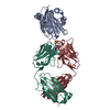
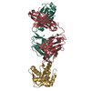
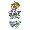
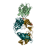
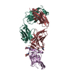
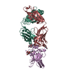
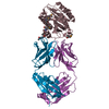
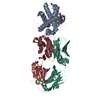
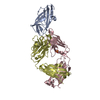
| Title | Crystal structure of Human Angiogenin in complex with Fab fragment of its monoclonal antibody mAb 26-2F | |||||||||
 Components Components |
| |||||||||
 Keywords Keywords | IMMUNE SYSTEM/HYDROLASE / COMPLEX (ANTIBODY-HYDROLASE) / RIBONUCLEASE / ANTIBODY / IMMUNE SYSTEM-HYDROLASE complex | |||||||||
| Function / homology |  Function and homology information Function and homology informationangiogenin-PRI complex / tRNA-specific ribonuclease activity / negative regulation of translation in response to stress / tRNA-derived small RNA (tsRNA or tRNA-related fragment, tRF) biogenesis / tRNA decay / signaling / cell communication / Hydrolases; Acting on ester bonds; Endoribonucleases producing 3'-phosphomonoesters / Adherens junctions interactions / oocyte maturation ...angiogenin-PRI complex / tRNA-specific ribonuclease activity / negative regulation of translation in response to stress / tRNA-derived small RNA (tsRNA or tRNA-related fragment, tRF) biogenesis / tRNA decay / signaling / cell communication / Hydrolases; Acting on ester bonds; Endoribonucleases producing 3'-phosphomonoesters / Adherens junctions interactions / oocyte maturation / hematopoietic stem cell proliferation / homeostatic process / rRNA transcription / basement membrane / positive regulation of phosphorylation / RNA nuclease activity / endocytic vesicle / ovarian follicle development / actin filament polymerization / peptide binding / RNA endonuclease activity / response to hormone / positive regulation of endothelial cell proliferation / placenta development / stress granule assembly / positive regulation of protein secretion / negative regulation of smooth muscle cell proliferation / cytoplasmic stress granule / antimicrobial humoral immune response mediated by antimicrobial peptide / cell migration / heparin binding / chromosome / antibacterial humoral response / actin cytoskeleton / growth cone / ribosome binding / actin binding / endonuclease activity / angiogenesis / response to hypoxia / rRNA binding / defense response to Gram-positive bacterium / receptor ligand activity / copper ion binding / signaling receptor binding / innate immune response / hydrolase activity / neuronal cell body / negative regulation of apoptotic process / nucleolus / signal transduction / protein homodimerization activity / : / DNA binding / extracellular region / nucleus / cytoplasm / cytosol Similarity search - Function | |||||||||
| Biological species |  HOMO SAPIENS (human) HOMO SAPIENS (human) | |||||||||
| Method |  X-RAY DIFFRACTION / SYNCHROTRON / MOLECULAR REPLACEMENT / Resolution: 2 Å X-RAY DIFFRACTION / SYNCHROTRON / MOLECULAR REPLACEMENT / Resolution: 2 Å | |||||||||
 Authors Authors | Chavali, G.B. / Papageorgiou, A.C. / Acharya, K.R. | |||||||||
 Citation Citation | Journal: Structure / Year: 2003 Title: The Crystal Structure of Human Angiogenin in Complex with an Antitumor Neutralizing Antibody Authors: Chavali, G.B. / Papageorgiou, A.C. / Olson, K. / Fett, J. / Hu, G. / Shapiro, R. / Acharya, K.R. | |||||||||
| History |
| |||||||||
| Remark 700 | SHEET THE SHEET STRUCTURE OF THIS MOLECULE IS BIFURCATED. IN ORDER TO REPRESENT THIS FEATURE IN ... SHEET THE SHEET STRUCTURE OF THIS MOLECULE IS BIFURCATED. IN ORDER TO REPRESENT THIS FEATURE IN THE SHEET RECORDS BELOW, TWO SHEETS ARE DEFINED. |
- Structure visualization
| Structure viewer | Molecule: MolmilJmol/JSmol |
|---|
- Downloads & links
-Download
| PDBx/mmCIF format | 1h0d.cif.gz | 131.1 KB | Display | PDBx/mmCIF format |
|---|---|---|---|---|
| PDB format | pdb1h0d.ent.gz | 100.5 KB | Display | PDB format |
| PDBx/mmJSON format | 1h0d.json.gz | Tree view | PDBx/mmJSON format | |
| Others |  Other downloads Other downloads |
-Validation report
| Arichive directory | https://data.pdbj.org/pub/pdb/validation_reports/h0/1h0dftp://data.pdbj.org/pub/pdb/validation_reports/h0/1h0d | HTTPS FTP |
|---|
-Related structure data
| Related structure data | 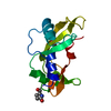 1b1iS 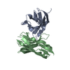 1fvcS 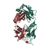 1tetS  3hfl S: Starting model for refinement |
|---|---|
| Similar structure data |

-Links
 PDBj
PDBj


- Assembly
| Deposited unit | 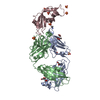
| ||||||||
|---|---|---|---|---|---|---|---|---|---|
| 1 |
| ||||||||
| Unit cell |
| ||||||||
| Components on special symmetry positions |
|
-Components
-Protein , 1 types, 1 molecules C
| #3: Protein | Mass: 14152.006 Da / Num. of mol.: 1 Source method: isolated from a genetically manipulated source Source: (gene. exp.) HOMO SAPIENS (human) / Production host:  |
|---|
-Antibody , 2 types, 2 molecules AB
| #1: Antibody | Mass: 23584.064 Da / Num. of mol.: 1 Source method: isolated from a genetically manipulated source Details: HINGE REGION OBSERVED IN THE FAB FRAGMENT / Source: (gene. exp.) HOMO SAPIENS (human) / Production host: |
|---|---|
| #2: Antibody | Mass: 23658.602 Da / Num. of mol.: 1 Source method: isolated from a genetically manipulated source Details: HINGE REGION OBSERVED IN THE FAB FRAGMENT / Source: (gene. exp.) HOMO SAPIENS (human) / Production host: |
-Non-polymers , 3 types, 341 molecules 


| #4: Chemical | ChemComp-SO4 /  Mass: 96.063 Da / Num. of mol.: 8 / Source method: obtained synthetically / Formula: SO4 Mass: 96.063 Da / Num. of mol.: 8 / Source method: obtained synthetically / Formula: SO4#5: Chemical | ChemComp-GOL /  Mass: 92.094 Da / Num. of mol.: 6 / Source method: obtained synthetically / Formula: C3H8O3 Mass: 92.094 Da / Num. of mol.: 6 / Source method: obtained synthetically / Formula: C3H8O3#6: Water | ChemComp-HOH / | Mass: 18.015 Da / Num. of mol.: 327 / Source method: isolated from a natural source / Formula: H2O |
|---|
-Details
| Compound details | THERE ARE CHANGES IN RESIDUES FOR CHAIN A AND B (LYS146ARG,GLU165GL AND IN CHAIN B ARG189TRP) WITH ...THERE ARE CHANGES IN RESIDUES FOR CHAIN A AND B (LYS146ARG,GLU165GL AND IN CHAIN B ARG189TRP) WITH RESPECT TO THE KABAT DATABASE SEQUENCES FOR FAB CONSTANT REGION. |
|---|---|
| Has protein modification | Y |
| Sequence details | RESIDUES 128-133 OF CHAIN B WERE MODELLED AS GLYCINES. RESIDUES 198,199,210,211 OF CHAIN A WERE ...RESIDUES 128-133 OF CHAIN B WERE MODELLED AS GLYCINES. RESIDUES 198,199,210,211 OF CHAIN A WERE MODELLED AS ALANINES RESIDUES 134,213,214,215 OF CHAIN B WERE MODELLED AS ALANINES RESIDUES 1 OF CHAIN C WAS MODELLED AS ALANINE |
-Experimental details
-Experiment
| Experiment | Method: X-RAY DIFFRACTION / Number of used crystals: 1 |
|---|
- Sample preparation
| Crystal | Density Matthews: 2.86 Å3/Da / Density % sol: 57.04 % | ||||||||||||||||||||||||||||
|---|---|---|---|---|---|---|---|---|---|---|---|---|---|---|---|---|---|---|---|---|---|---|---|---|---|---|---|---|---|
| Crystal grow | pH: 6 / Details: 30% PEG 3350 AND 0.2M LITHIUM SULPHATE PH 6.0-7.5 | ||||||||||||||||||||||||||||
| Crystal grow | *PLUS Temperature: 16 ℃ / pH: 7.2 / Method: vapor diffusion, hanging drop | ||||||||||||||||||||||||||||
| Components of the solutions | *PLUS
|
-Data collection
| Diffraction | Mean temperature: 100 K |
|---|---|
| Diffraction source | Source: SYNCHROTRON / Site: SRS  / Beamline: PX9.6 / Wavelength: 0.87 / Beamline: PX9.6 / Wavelength: 0.87 |
| Detector | Type: ADSC CCD / Detector: CCD / Date: Apr 15, 2000 |
| Radiation | Protocol: SINGLE WAVELENGTH / Monochromatic (M) / Laue (L): M / Scattering type: x-ray |
| Radiation wavelength | Wavelength: 0.87 Å / Relative weight: 1 |
| Reflection | Resolution: 2→40 Å / Num. obs: 46470 / % possible obs: 98.9 % / Redundancy: 3 % / Biso Wilson estimate: 17.5 Å2 / Rmerge(I) obs: 0.061 / Net I/σ(I): 17.6 |
| Reflection shell | Resolution: 2→2.07 Å / Rmerge(I) obs: 0.2 / Mean I/σ(I) obs: 4.92 / % possible all: 97.8 |
| Reflection | *PLUS Highest resolution: 2 Å / Lowest resolution: 40 Å / Rmerge(I) obs: 0.061 |
| Reflection shell | *PLUS % possible obs: 97.8 % / Rmerge(I) obs: 0.2 / Mean I/σ(I) obs: 4.92 |
- Processing
| Software |
| ||||||||||||||||||||||||||||||||||||||||||||||||||||||||||||
|---|---|---|---|---|---|---|---|---|---|---|---|---|---|---|---|---|---|---|---|---|---|---|---|---|---|---|---|---|---|---|---|---|---|---|---|---|---|---|---|---|---|---|---|---|---|---|---|---|---|---|---|---|---|---|---|---|---|---|---|---|---|
| Refinement | Method to determine structure: MOLECULAR REPLACEMENT Starting model: PDB ENTRIES 1FVC,3HFL,1TET,1B1I Resolution: 2→40 Å / Rfactor Rfree error: 0.006 / Isotropic thermal model: RESTRAINED / Cross valid method: THROUGHOUT / σ(F): 0
| ||||||||||||||||||||||||||||||||||||||||||||||||||||||||||||
| Displacement parameters | Biso mean: 39.7 Å2
| ||||||||||||||||||||||||||||||||||||||||||||||||||||||||||||
| Refine analyze |
| ||||||||||||||||||||||||||||||||||||||||||||||||||||||||||||
| Refinement step | Cycle: LAST / Resolution: 2→40 Å
| ||||||||||||||||||||||||||||||||||||||||||||||||||||||||||||
| Refine LS restraints |
| ||||||||||||||||||||||||||||||||||||||||||||||||||||||||||||
| LS refinement shell | Resolution: 2→2.13 Å / Rfactor Rfree error: 0.022 / Total num. of bins used: 6
| ||||||||||||||||||||||||||||||||||||||||||||||||||||||||||||
| Xplor file |
| ||||||||||||||||||||||||||||||||||||||||||||||||||||||||||||
| Refinement | *PLUS Lowest resolution: 40 Å / % reflection Rfree: 5 % | ||||||||||||||||||||||||||||||||||||||||||||||||||||||||||||
| Solvent computation | *PLUS | ||||||||||||||||||||||||||||||||||||||||||||||||||||||||||||
| Displacement parameters | *PLUS | ||||||||||||||||||||||||||||||||||||||||||||||||||||||||||||
| Refine LS restraints | *PLUS
|
