SHEET DETERMINATION METHOD: DSSP THE SHEETS PRESENTED AS "AA" AND "BA" IN EACH CHAIN ON SHEET ... SHEET DETERMINATION METHOD: DSSP THE SHEETS PRESENTED AS "AA" AND "BA" IN EACH CHAIN ON SHEET RECORDS BELOW ARE ACTUALLY 9-STRANDED BARRELS THESE ARE REPRESENTED BY A 10-STRANDED SHEET IN WHICH THE FIRST AND LAST STRANDS ARE IDENTICAL.
Mass: 18.015 Da / Num. of mol.: 389 / Source method: isolated from a natural source / Formula: H2O
Has protein modification
Y
-
Experimental details
-
Experiment
Experiment
Method: X-RAY DIFFRACTION
-
Sample preparation
Crystal
Density Matthews: 2.19 Å3/Da / Density % sol: 43.75 % Description: DUE TO LOW COMPLETENESS AT LOW RESOLUTION, THE MISSING LOW RESOLUTION REFLECTIONS WERE 'REFILLED' WITH DATA FROM AN IN HOUSE COLLECTION AFTER THE TWO DATA SETS WERE SCALED TOGETHER. THE ...Description: DUE TO LOW COMPLETENESS AT LOW RESOLUTION, THE MISSING LOW RESOLUTION REFLECTIONS WERE 'REFILLED' WITH DATA FROM AN IN HOUSE COLLECTION AFTER THE TWO DATA SETS WERE SCALED TOGETHER. THE STATISTICS REPORTED REFER TO THE SYNCHROTRON DATA SET. AFTER REFILLING THE OVERALL COMPLETENESS WAS 88.5% (99.2% BETWEEN 35 AND 5 A RESOLUTION)
Crystal grow
Method: vapor diffusion, hanging drop / pH: 9 Details: HANGING DROP CONSISTING OF 2 UL OF PROTEIN (31.2 MG/ML) AND 2 UL RESERVOIR (0.1 M TRIS-HCL PH9.0, 1.6 M AMMONIUM SULPHATE), pH 9.00
Crystal grow
*PLUS
Temperature: 291 K / pH: 8.5 / Method: vapor diffusion, hanging drop Details: Loleggio, L., (1997) Acta Crystallogr.,Sect.D, 53, 599.
In the structure databanks used in Yorodumi, some data are registered as the other names, "COVID-19 virus" and "2019-nCoV". Here are the details of the virus and the list of structure data.
Jan 31, 2019. EMDB accession codes are about to change! (news from PDBe EMDB page)
EMDB accession codes are about to change! (news from PDBe EMDB page)
The allocation of 4 digits for EMDB accession codes will soon come to an end. Whilst these codes will remain in use, new EMDB accession codes will include an additional digit and will expand incrementally as the available range of codes is exhausted. The current 4-digit format prefixed with “EMD-” (i.e. EMD-XXXX) will advance to a 5-digit format (i.e. EMD-XXXXX), and so on. It is currently estimated that the 4-digit codes will be depleted around Spring 2019, at which point the 5-digit format will come into force.
The EM Navigator/Yorodumi systems omit the EMD- prefix.
Related info.:Q: What is EMD? / ID/Accession-code notation in Yorodumi/EM Navigator
Yorodumi is a browser for structure data from EMDB, PDB, SASBDB, etc.
This page is also the successor to EM Navigator detail page, and also detail information page/front-end page for Omokage search.
The word "yorodu" (or yorozu) is an old Japanese word meaning "ten thousand". "mi" (miru) is to see.
Related info.:EMDB / PDB / SASBDB / Comparison of 3 databanks / Yorodumi Search / Aug 31, 2016. New EM Navigator & Yorodumi / Yorodumi Papers / Jmol/JSmol / Function and homology information / Changes in new EM Navigator and Yorodumi
 Movie
Movie Controller


 Open data
 Basic information
 Components
 Keywords
 Function and homology information
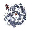
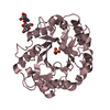
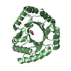
 Thermoascus aurantiacus (fungus)
 X-RAY DIFFRACTION /
  Authors
 Citation
 Structure visualization
 Downloads & links
 Other downloads
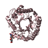


 PDBj
PDBj Assembly


 Mass: 18.015 Da / Num. of mol.: 389 / Source method: isolated from a natural source / Formula: H2O
 Sample preparation
 / Beamline: BL-6A / Wavelength: 1
  Processing