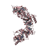
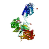
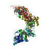
High-molecular-weight cytochrome c / : / : / Outer membrane cytochrome MtrC/MtrF-like, domains II/IV / : / Class III cytochrome C / Class III cytochrome C family / Cytochrome c, class III / Cytochrome C3 / Cytochrome C3 ...High-molecular-weight cytochrome c / : / : / Outer membrane cytochrome MtrC/MtrF-like, domains II/IV / : / Class III cytochrome C / Class III cytochrome C family / Cytochrome c, class III / Cytochrome C3 / Cytochrome C3 / Multiheme cytochrome c family profile. / Multiheme cytochrome superfamily / Alpha-Beta Complex / Alpha Beta Similarity search - Domain/homology
Resolution: 2.5→27 Å / Num. obs: 23044 / % possible obs: 97.3 % / Redundancy: 5.2 % / Rmerge(I) obs: 0.071 / Net I/σ(I): 7.3
Reflection shell
Resolution: 2.5→2.59 Å / Redundancy: 3.1 % / Rmerge(I) obs: 0.421 / Mean I/σ(I) obs: 1.9 / % possible all: 84.6
Reflection
*PLUS
Highest resolution: 2.42 Å / Lowest resolution: 27 Å / % possible obs: 99.4 % / Redundancy: 7.6 % / Rmerge(I) obs: 0.061
Reflection shell
*PLUS
% possible obs: 99.2 % / Redundancy: 7.5 % / Rmerge(I) obs: 0.331 / Mean I/σ(I) obs: 1.5
-
Processing
Software
Name
Version
Classification
REFMAC
5.0.36
refinement
DENZO
datareduction
SCALA
datascaling
SHARP
phasing
Refinement
Method to determine structure: MAD / Resolution: 2.4→33.52 Å / Cor.coef. Fo:Fc: 0.947 / Cor.coef. Fo:Fc free: 0.909 / SU B: 9.016 / SU ML: 0.215 / TLS residual ADP flag: LIKELY RESIDUAL / Cross valid method: THROUGHOUT / ESU R: 0.407 / ESU R Free: 0.288 / Stereochemistry target values: MAXIMUM LIKELIHOOD Details: ALA A 144, NO DENSITY PRESENT FOR SIDE CHAIN, THE RESIDUE WAS THERFORE MODELED AS ALA ALA A 512, NO DENSITY PRESENT FOR SIDE CHAIN, THE RESIDUE WAS THERFORE MODELED AS ALA
Rfactor
Num. reflection
% reflection
Selection details
Rfree
0.276
1281
4.8 %
RANDOM
Rwork
0.201
-
-
-
obs
-
25316
100 %
-
Solvent computation
Ion probe radii: 0.8 Å / Shrinkage radii: 0.8 Å / VDW probe radii: 1.4 Å / Solvent model: BABINET MODEL PLUS MASK
 Movie
Movie Controller

 Yorodumi
 Open data
 Basic information
 Components
 Keywords
 Function and homology information
 DESULFOVIBRIO VULGARIS (bacteria)
 X-RAY DIFFRACTION /
  Authors
 Citation
 Structure visualization
 Downloads & links
 Other downloads
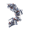

 PDBj
PDBj


 Assembly
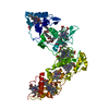

 Mass: 618.503 Da / Num. of mol.: 16 / Source method: obtained synthetically / Formula: C34H34FeN4O4
 Mass: 18.015 Da / Num. of mol.: 151 / Source method: isolated from a natural source / Formula: H2O
 Sample preparation
 / Beamline: ID29 / Wavelength: 1.73891
  Processing