 Movie
Movie Controller


+ Open data

- Basic information
| Entry | Database: PDB / ID: 1gsh | ||||||
|---|---|---|---|---|---|---|---|
| Title | STRUCTURE OF ESCHERICHIA COLI GLUTATHIONE SYNTHETASE AT PH 7.5 | ||||||
 Components Components | GLUTATHIONE BIOSYNTHETIC LIGASE | ||||||
 Keywords Keywords | GLUTATHIONE BIOSYNTHESIS LIGASE / GLUTATHIONE BIOSYNTHESIS / GLUTATHIONE SYNTHASE | ||||||
| Function / homology |  Function and homology information Function and homology informationglutathione synthase / glutathione synthase activity / glutathione biosynthetic process / protein homotetramerization / magnesium ion binding / ATP binding / identical protein binding / cytoplasm / cytosol Similarity search - Function | ||||||
| Biological species |  | ||||||
| Method |  X-RAY DIFFRACTION / SYNCHROTRON / Resolution: 2 Å X-RAY DIFFRACTION / SYNCHROTRON / Resolution: 2 Å | ||||||
 Authors Authors | Matsuda, K. / Kato, H. / Yamaguchi, H. / Nishioka, T. / Katsube, Y. / Oda, J. | ||||||
 Citation Citation | Journal: Protein Eng. / Year: 1996 Title: Crystal structure of glutathione synthetase at optimal pH: domain architecture and structural similarity with other proteins. Authors: Matsuda, K. / Mizuguchi, K. / Nishioka, T. / Kato, H. / Go, N. / Oda, J. #1: Journal: Biochemistry / Year: 1994Title: Flexible Loop that is Novel Catalytic Machinery in a Ligase. Atomic Structure and Function of the Loopless Glutathione Synthetase Authors: Kato, H. / Tanaka, T. / Yamaguchi, H. / Hara, T. / Nishioka, T. / Katsube, Y. / Oda, J. #2: Journal: J.Am.Chem.Soc. / Year: 1994Title: Mechanism-Based Inactivation of Glutathione Synthetase by Phosphinic Acid Transition-State Analogue Authors: Hiratake, J. / Kato, H. / Oda, J. #3: Journal: Biochemistry / Year: 1993Title: Use of Adenosine (5')Polyphospho(5')Pyridoxals to Study the Substrate-Binding Region of Glutathione Synthetase from Escherichia Coli B Authors: Hibi, T. / Kato, H. / Nishioka, T. / Oda, J. / Yamaguchi, H. / Katsube, Y. / Tanizawa, K. / Fukui, T. #4: Journal: Biochemistry / Year: 1993Title: Flexibility Impaired by Mutations Revealed the Multifunctional Roles of the Loop in Glutathione Synthetase Authors: Tanaka, T. / Yamaguchi, H. / Kato, H. / Nishioka, T. / Katsube, Y. / Oda, J. #5: Journal: J.Mol.Biol. / Year: 1993Title: Three-Dimensional Structure of the Glutathione Synthetase from Escherichia Coli B at 2.0 A Resolution Authors: Yamaguchi, H. / Kato, H. / Hata, Y. / Nishioka, T. / Kimura, A. / Oda, J. / Katsube, Y. #6: Journal: Photon Factory Activity Report / Year: 1992Title: Structural Studies on Glutathione Synthetase from Escherichia Coli B Authors: Yamaguchi, H. / Kato, H. / Hata, Y. / Nishioka, T. / Oda, J. / Katsube, Y. #7: Journal: J.Mol.Biol. / Year: 1989Title: Crystallization and Preliminary X-Ray Studies of Glutathione Synthetase from Escherichia Coli B Authors: Kato, H. / Yamaguchi, H. / Hata, Y. / Nishioka, T. / Katsube, Y. / Oda, J. | ||||||
| History |
|
- Structure visualization
| Structure viewer | Molecule: MolmilJmol/JSmol |
|---|
- Downloads & links
-Download
| PDBx/mmCIF format | 1gsh.cif.gz | 73.3 KB | Display | PDBx/mmCIF format |
|---|---|---|---|---|
| PDB format | pdb1gsh.ent.gz | 55.1 KB | Display | PDB format |
| PDBx/mmJSON format | 1gsh.json.gz | Tree view | PDBx/mmJSON format | |
| Others |  Other downloads Other downloads |
-Validation report
| Arichive directory | https://data.pdbj.org/pub/pdb/validation_reports/gs/1gshftp://data.pdbj.org/pub/pdb/validation_reports/gs/1gsh | HTTPS FTP |
|---|
-Related structure data


-Links
 PDBj
PDBj


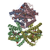
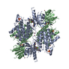
- Assembly
| Deposited unit | 
| ||||||||
|---|---|---|---|---|---|---|---|---|---|
| 1 | 
| ||||||||
| Unit cell |
|
-Components
| #1: Protein | Mass: 35601.824 Da / Num. of mol.: 1 Source method: isolated from a genetically manipulated source Source: (gene. exp.) |
|---|---|
| #2: Water | ChemComp-HOH /  Mass: 18.015 Da / Num. of mol.: 96 / Source method: isolated from a natural source / Formula: H2O Mass: 18.015 Da / Num. of mol.: 96 / Source method: isolated from a natural source / Formula: H2O |
-Experimental details
-Experiment
| Experiment | Method: X-RAY DIFFRACTION |
|---|
- Sample preparation
| Crystal | Density Matthews: 2.81 Å3/Da / Density % sol: 44 % | ||||||||||||||||||||||||
|---|---|---|---|---|---|---|---|---|---|---|---|---|---|---|---|---|---|---|---|---|---|---|---|---|---|
| Crystal grow | Method: microdialysis / pH: 7.5 Details: CRYSTALS WERE PREPARED AT PH 7.5 BY MICRODIALYSIS METHOD WITH AMMONIUM SULFATE AS THE PRECIPITATING AGENT. THE INNER SOLUTION 100 MICROLITER CONTAINED 1.5 % (W/V) GSHASE, 5 MM MGCL2 AND 10 % ...Details: CRYSTALS WERE PREPARED AT PH 7.5 BY MICRODIALYSIS METHOD WITH AMMONIUM SULFATE AS THE PRECIPITATING AGENT. THE INNER SOLUTION 100 MICROLITER CONTAINED 1.5 % (W/V) GSHASE, 5 MM MGCL2 AND 10 % SATURATED AMMONIUM SULFATE IN 50 MM TRIS-HCL BUFFER (PH 7.5). THE INNER SOLUTION WAS DIALYZED AGAINST 50 MM TRIS-HCL BUFFER (PH 7.5) WHICH CONTAINED 5 MM MGCL2 AND 25 % SATURATED AMMONIUM SULFATE.TRIS-HCL BUFFER, microdialysis | ||||||||||||||||||||||||
| Crystal | *PLUS | ||||||||||||||||||||||||
| Crystal grow | *PLUS Method: unknown | ||||||||||||||||||||||||
| Components of the solutions | *PLUS
|
-Data collection
| Diffraction source | Source: SYNCHROTRON / Site: Photon Factory  / Beamline: BL-6A / Wavelength: 1.04 / Beamline: BL-6A / Wavelength: 1.04 |
|---|---|
| Detector | Type: RIGAKU / Detector: IMAGE PLATE / Date: Mar 18, 1991 |
| Radiation | Monochromatic (M) / Laue (L): M / Scattering type: x-ray |
| Radiation wavelength | Wavelength: 1.04 Å / Relative weight: 1 |
| Reflection | Highest resolution: 2 Å / Num. obs: 30967 / % possible obs: 90.3 % / Observed criterion σ(I): 1 / Rmerge(I) obs: 0.07 |
| Reflection | *PLUS Highest resolution: 1.8 Å / % possible obs: 84.1 % / Rmerge(I) obs: 0.0701 |
- Processing
| Software |
| ||||||||||||||||||||||||||||||||||||||||||||||||||||||||||||
|---|---|---|---|---|---|---|---|---|---|---|---|---|---|---|---|---|---|---|---|---|---|---|---|---|---|---|---|---|---|---|---|---|---|---|---|---|---|---|---|---|---|---|---|---|---|---|---|---|---|---|---|---|---|---|---|---|---|---|---|---|---|
| Refinement | Resolution: 2→10 Å / σ(F): 2
| ||||||||||||||||||||||||||||||||||||||||||||||||||||||||||||
| Displacement parameters | Biso mean: 18.2 Å2 | ||||||||||||||||||||||||||||||||||||||||||||||||||||||||||||
| Refine analyze | Luzzati coordinate error obs: 0.18 Å | ||||||||||||||||||||||||||||||||||||||||||||||||||||||||||||
| Refinement step | Cycle: LAST / Resolution: 2→10 Å
| ||||||||||||||||||||||||||||||||||||||||||||||||||||||||||||
| Refine LS restraints |
| ||||||||||||||||||||||||||||||||||||||||||||||||||||||||||||
| Xplor file |
| ||||||||||||||||||||||||||||||||||||||||||||||||||||||||||||
| Software | *PLUS Name: X-PLOR / Version: 3.1 / Classification: refinement | ||||||||||||||||||||||||||||||||||||||||||||||||||||||||||||
| Refinement | *PLUS % reflection Rfree: 10 % / Rfactor Rfree: 23.5 | ||||||||||||||||||||||||||||||||||||||||||||||||||||||||||||
| Solvent computation | *PLUS | ||||||||||||||||||||||||||||||||||||||||||||||||||||||||||||
| Displacement parameters | *PLUS | ||||||||||||||||||||||||||||||||||||||||||||||||||||||||||||
| Refine LS restraints | *PLUS Type: x_dihedral_angle_d / Dev ideal: 25.359 |
