 Movie
Movie Controller


+ Open data

- Basic information
| Entry | Database: PDB / ID: 1gmx | ||||||
|---|---|---|---|---|---|---|---|
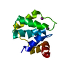
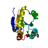
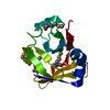
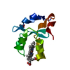
| Title | Escherichia coli GlpE sulfurtransferase | ||||||
 Components Components | THIOSULFATE SULFURTRANSFERASE GLPE | ||||||
 Keywords Keywords | TRANSFERASE / RHODANESE / SULFURTRANSFERASE / GLYCEROL METABOLISM | ||||||
| Function / homology |  Function and homology information Function and homology informationsulfur utilization / thiosulfate-thioredoxin sulfurtransferase activity / thiosulfate sulfurtransferase / thiosulfate-cyanide sulfurtransferase activity / cytoplasm Similarity search - Function | ||||||
| Biological species |  | ||||||
| Method |  X-RAY DIFFRACTION / SYNCHROTRON / SIRAS / Resolution: 1.1 Å X-RAY DIFFRACTION / SYNCHROTRON / SIRAS / Resolution: 1.1 Å | ||||||
 Authors Authors | Spallarossa, A. / Donahue, J.T. / Larson, T.J. / Bolognesi, M. / Bordo, D. | ||||||
 Citation Citation | Journal: Structure / Year: 2001 Title: Escherichia Coli Glpe is a Prototype Sulfurtransferase for the Single-Domain Rhodanese Homology Superfamily Authors: Spallarossa, A. / Donahue, J.T. / Larson, T.J. / Bolognesi, M. / Bordo, D. | ||||||
| History |
|
- Structure visualization
| Structure viewer | Molecule: MolmilJmol/JSmol |
|---|
- Downloads & links
-Download
| PDBx/mmCIF format | 1gmx.cif.gz | 61.6 KB | Display | PDBx/mmCIF format |
|---|---|---|---|---|
| PDB format | pdb1gmx.ent.gz | 45.1 KB | Display | PDB format |
| PDBx/mmJSON format | 1gmx.json.gz | Tree view | PDBx/mmJSON format | |
| Others |  Other downloads Other downloads |
-Validation report
| Arichive directory | https://data.pdbj.org/pub/pdb/validation_reports/gm/1gmxftp://data.pdbj.org/pub/pdb/validation_reports/gm/1gmx | HTTPS FTP |
|---|
-Related structure data


-Links
 PDBj
PDBj- Assembly
| Deposited unit | 
| ||||||||
|---|---|---|---|---|---|---|---|---|---|
| 1 |
| ||||||||
| Unit cell |
|
-Components
| #1: Protein | Mass: 12124.485 Da / Num. of mol.: 1 Source method: isolated from a genetically manipulated source Source: (gene. exp.) | ||||||
|---|---|---|---|---|---|---|---|
| #2: Chemical |   Mass: 59.044 Da / Num. of mol.: 2 / Source method: obtained synthetically / Formula: C2H3O2 Mass: 59.044 Da / Num. of mol.: 2 / Source method: obtained synthetically / Formula: C2H3O2#3: Chemical | ChemComp-EDO /   Mass: 62.068 Da / Num. of mol.: 7 / Source method: obtained synthetically / Formula: C2H6O2 Mass: 62.068 Da / Num. of mol.: 7 / Source method: obtained synthetically / Formula: C2H6O2#4: Water | ChemComp-HOH / |  Mass: 18.015 Da / Num. of mol.: 98 / Source method: isolated from a natural source / Formula: H2O Mass: 18.015 Da / Num. of mol.: 98 / Source method: isolated from a natural source / Formula: H2OHas protein modification | Y | |
-Experimental details
-Experiment
| Experiment | Method: X-RAY DIFFRACTION / Number of used crystals: 1 |
|---|
- Sample preparation
| Crystal | Density Matthews: 2.1 Å3/Da / Density % sol: 42 % | |||||||||||||||||||||||||||||||||||||||||||||||||||||||||||||||
|---|---|---|---|---|---|---|---|---|---|---|---|---|---|---|---|---|---|---|---|---|---|---|---|---|---|---|---|---|---|---|---|---|---|---|---|---|---|---|---|---|---|---|---|---|---|---|---|---|---|---|---|---|---|---|---|---|---|---|---|---|---|---|---|---|
| Crystal grow | pH: 4.6 / Details: pH 4.60 | |||||||||||||||||||||||||||||||||||||||||||||||||||||||||||||||
| Crystal grow | *PLUS Method: vapor diffusion, hanging drop | |||||||||||||||||||||||||||||||||||||||||||||||||||||||||||||||
| Components of the solutions | *PLUS
|
-Data collection
| Diffraction | Mean temperature: 100 K |
|---|---|
| Diffraction source | Source: SYNCHROTRON / Site: EMBL/DESY, HAMBURG  / Beamline: X11 / Wavelength: 0.91 / Beamline: X11 / Wavelength: 0.91 |
| Detector | Date: Oct 15, 2000 |
| Radiation | Protocol: SINGLE WAVELENGTH / Monochromatic (M) / Laue (L): M / Scattering type: x-ray |
| Radiation wavelength | Wavelength: 0.91 Å / Relative weight: 1 |
| Reflection | Resolution: 1.06→20 Å / Num. obs: 45040 / % possible obs: 99.6 % / Observed criterion σ(I): 3 / Redundancy: 1.2 % / Rmerge(I) obs: 0.023 / Net I/σ(I): 43 |
| Reflection shell | Resolution: 1.06→1.08 Å / Rmerge(I) obs: 0.281 / Mean I/σ(I) obs: 3 / % possible all: 93.6 |
| Reflection | *PLUS Lowest resolution: 20 Å / Num. measured all: 555366 |
| Reflection shell | *PLUS % possible obs: 93.6 % |
- Processing
| Software |
| ||||||||||||||||||||
|---|---|---|---|---|---|---|---|---|---|---|---|---|---|---|---|---|---|---|---|---|---|
| Refinement | Method to determine structure: SIRAS / Resolution: 1.1→47 Å / SU B: 0.75 / SU ML: 0.02 / Cross valid method: THROUGHOUT / ESU R Free: 0.03
| ||||||||||||||||||||
| Refinement step | Cycle: LAST / Resolution: 1.1→47 Å
| ||||||||||||||||||||
| Refinement | *PLUS Highest resolution: 1.06 Å / Lowest resolution: 47 Å / % reflection Rfree: 5 % / Rfactor obs: 0.128 / Rfactor Rfree: 0.151 / Rfactor Rwork: 0.128 | ||||||||||||||||||||
| Solvent computation | *PLUS | ||||||||||||||||||||
| Displacement parameters | *PLUS | ||||||||||||||||||||
| Refine LS restraints | *PLUS
|
