 Movie
Movie Controller

[English] 日本語
 Yorodumi
- PDB-1gmj: The structure of bovine IF1, the regulatory subunit of mitochondr... -
+ Open data

- Basic information
| Entry | Database: PDB / ID: 1gmj | ||||||
|---|---|---|---|---|---|---|---|
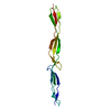
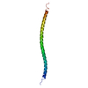
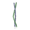
| Title | The structure of bovine IF1, the regulatory subunit of mitochondrial F-ATPase | ||||||
 Components Components | ATPASE INHIBITOR | ||||||
 Keywords Keywords | ATPASE INHIBITOR / BOVINE F1-ATPASE INHIBITOR PROTEIN / COILED-COIL STRUCTURE / P DEPENDENT OLIGOMERIZATION / ATP HYDROLYSIS | ||||||
| Function / homology |  Function and homology information Function and homology informationnegative regulation of mitochondrial ATP synthesis coupled proton transport / angiostatin binding / negative regulation of hydrolase activity / ATPase inhibitor activity / heme biosynthetic process / negative regulation of endothelial cell proliferation / erythrocyte differentiation / ATPase binding / protein homotetramerization / calmodulin binding ...negative regulation of mitochondrial ATP synthesis coupled proton transport / angiostatin binding / negative regulation of hydrolase activity / ATPase inhibitor activity / heme biosynthetic process / negative regulation of endothelial cell proliferation / erythrocyte differentiation / ATPase binding / protein homotetramerization / calmodulin binding / structural molecule activity / cell surface / protein homodimerization activity / protein-containing complex / mitochondrion / identical protein binding / cytoplasm Similarity search - Function | ||||||
| Biological species |  | ||||||
| Method |  X-RAY DIFFRACTION / SYNCHROTRON / SIRAS / Resolution: 2.2 Å X-RAY DIFFRACTION / SYNCHROTRON / SIRAS / Resolution: 2.2 Å | ||||||
 Authors Authors | Cabezon, E. / Runswick, M.J. / Leslie, A.G.W. / Walker, J.E. | ||||||
 Citation Citation | Journal: Embo J. / Year: 2001 Title: The Structure of Bovine If(1), the Regulatory Subunit of Mitochondrial F-ATPase. Authors: Cabezon, E. / Runswick, M.J. / Leslie, A.G.W. / Walker, J.E. #1: Journal: J.Biol.Chem. / Year: 2000 Title: Modulation of the Oligomerization State of Bovine F1-ATPase Inhibitor Protein, If1, by Ph Authors: Cabezon, E. / Butler, P.J.G. / Runswick, M.J. / Walker, J.E. #2: Journal: J.Biol.Chem. / Year: 2000 Title: Dimerization of Bovine F1-ATPase by Binding the Inhibitor Protein, If1 Authors: Cabezon, E. / Arechaga, I. / Butler, P.J.G. / Walker, J.E. | ||||||
| History |
|
- Structure visualization
| Structure viewer | Molecule: MolmilJmol/JSmol |
|---|
- Downloads & links
-Download
| PDBx/mmCIF format | 1gmj.cif.gz | 60.1 KB | Display | PDBx/mmCIF format |
|---|---|---|---|---|
| PDB format | pdb1gmj.ent.gz | 46.2 KB | Display | PDB format |
| PDBx/mmJSON format | 1gmj.json.gz | Tree view | PDBx/mmJSON format | |
| Others |  Other downloads Other downloads |
-Validation report
| Arichive directory | https://data.pdbj.org/pub/pdb/validation_reports/gm/1gmjftp://data.pdbj.org/pub/pdb/validation_reports/gm/1gmj | HTTPS FTP |
|---|
-Related structure data
| Related structure data | |
|---|---|
| Similar structure data |


-Links
 PDBj
PDBj- Assembly
| Deposited unit | 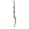
| ||||||||
|---|---|---|---|---|---|---|---|---|---|
| 1 | 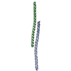
| ||||||||
| 2 | 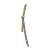
| ||||||||
| Unit cell |
| ||||||||
| Noncrystallographic symmetry (NCS) | NCS oper: (Code: given / Matrix: (1),| Details | THE ACTIVE FORM OF THE PROTEIN IS DIMERIC . IN THE CRYSTALTWO DIMERS INSTERACT TO FORM A DIMER OF DIMERS. | |
-Components
| #1: Protein | Mass: 9595.572 Da / Num. of mol.: 4 / Mutation: YES Source method: isolated from a genetically manipulated source Details: THE ACTIVE FORM OF THE PROTEIN IS DIMERIC (DIMERS AB OR CD) Source: (gene. exp.)  Compound details | CHAIN A, B, C, D ENGINEERED | Sequence details | SEQUENCE DATABASE CORRESPOND | |
|---|
-Experimental details
-Experiment
| Experiment | Method: X-RAY DIFFRACTION / Number of used crystals: 1 |
|---|
- Sample preparation
| Crystal | Density Matthews: 3.6 Å3/Da / Density % sol: 65 % Description: ANISOTROPIC DATA. WITHIN THE ANISOTROPIC RESOLUTION LIMITS USED FOR THE INTEGRATION, THE DATA SET IS 96% COMPLETE. | ||||||||||||||||||||||||||||||||||||
|---|---|---|---|---|---|---|---|---|---|---|---|---|---|---|---|---|---|---|---|---|---|---|---|---|---|---|---|---|---|---|---|---|---|---|---|---|---|
| Crystal grow | Temperature: 296 K / Method: vapor diffusion, sitting drop / pH: 8 Details: CRYSTALS WERE GROWN BY EQUILIBRATING A IF1-H49K SOLUTION, AT 6 MG/ML IN BUFFER 10 MM TRIS-HCL PH 8.0, RESERVOIR CONTAINING 0.8 M MONO-SODIUM DIHYDROGEN PHOSPHAT AGAINST A 0.8 M MONO- ...Details: CRYSTALS WERE GROWN BY EQUILIBRATING A IF1-H49K SOLUTION, AT 6 MG/ML IN BUFFER 10 MM TRIS-HCL PH 8.0, RESERVOIR CONTAINING 0.8 M MONO-SODIUM DIHYDROGEN PHOSPHAT AGAINST A 0.8 M MONO-POTASSIUM DIHYDROGEN PHOSPHATE AND 0.1 M HEPES-NA BUFFER PH 8, AT 23C, IN SITTING-DROP VAPOR-DIFFUSION TRAYS. | ||||||||||||||||||||||||||||||||||||
| Crystal grow | *PLUS Temperature: 23 ℃ / Method: vapor diffusion, sitting drop | ||||||||||||||||||||||||||||||||||||
| Components of the solutions | *PLUS
|
-Data collection
| Diffraction | Mean temperature: 100 K |
|---|---|
| Diffraction source | Source: SYNCHROTRON / Site: ESRF  / Beamline: ID14-1 / Wavelength: 0.934 / Beamline: ID14-1 / Wavelength: 0.934 |
| Detector | Type: MARRESEARCH / Detector: CCD / Date: Dec 15, 2000 |
| Radiation | Protocol: SINGLE WAVELENGTH / Monochromatic (M) / Laue (L): M / Scattering type: x-ray |
| Radiation wavelength | Wavelength: 0.934 Å / Relative weight: 1 |
| Reflection | Resolution: 2.2→14.2 Å / Num. obs: 18750 / % possible obs: 72.3 % / Redundancy: 2.4 % / Biso Wilson estimate: 33.1 Å2 / Rmerge(I) obs: 0.065 / Net I/σ(I): 10.5 |
| Reflection shell | Resolution: 2.2→2.35 Å / Redundancy: 2.4 % / Rmerge(I) obs: 0.227 / Mean I/σ(I) obs: 3.7 / % possible all: 10.8 |
| Reflection | *PLUS Highest resolution: 2.2 Å |
| Reflection shell | *PLUS % possible obs: 10.8 % / Redundancy: 2 % |
- Processing
| Software |
| ||||||||||||||||||||||||||||||||||||||||||||||||||||||||||||||||||||||||||||||||
|---|---|---|---|---|---|---|---|---|---|---|---|---|---|---|---|---|---|---|---|---|---|---|---|---|---|---|---|---|---|---|---|---|---|---|---|---|---|---|---|---|---|---|---|---|---|---|---|---|---|---|---|---|---|---|---|---|---|---|---|---|---|---|---|---|---|---|---|---|---|---|---|---|---|---|---|---|---|---|---|---|---|
| Refinement | Method to determine structure: SIRAS / Resolution: 2.2→26 Å / Rfactor Rfree error: 0.006 / Data cutoff high absF: 10000 / Isotropic thermal model: RESTRAINED / Cross valid method: THROUGHOUT / σ(F): 0 Stereochemistry target values: MAXIMUM LIKELIHOOD USING AMPLITUDES Details: 14 DIFFERENT GROUPS WERE USED IN THE NCS RESTRAINTS (TYPICAL WEIGHT 25, RMS 0.2,SIGB 10, RMSB 8) AN IN-HOUSE DATASET WAS USED TO PROVIDE LOW RESOLUTION STRUCTURE FACTORS (26-14A). . THERE ...Details: 14 DIFFERENT GROUPS WERE USED IN THE NCS RESTRAINTS (TYPICAL WEIGHT 25, RMS 0.2,SIGB 10, RMSB 8) AN IN-HOUSE DATASET WAS USED TO PROVIDE LOW RESOLUTION STRUCTURE FACTORS (26-14A). . THERE WAS NOT INTERPRETABLE ELECTRON DENSITY AT THE N-TERMINUS FOR RESIDUES 1-18; 1-19; 1-19 AND 1- 22 IN CHAINS A, B, C AND D, RESPECTIVELY AND FOR RESIDUES 84; 80-84; 79-84 AND 79-84 AT THE C-TERMINUS IN CHAINS A, B, C AND D, RESPECTIVELY.
| ||||||||||||||||||||||||||||||||||||||||||||||||||||||||||||||||||||||||||||||||
| Solvent computation | Solvent model: FLAT MODEL / Bsol: 53.1 Å2 / ksol: 0.4 e/Å3 | ||||||||||||||||||||||||||||||||||||||||||||||||||||||||||||||||||||||||||||||||
| Displacement parameters | Biso mean: 72.8 Å2
| ||||||||||||||||||||||||||||||||||||||||||||||||||||||||||||||||||||||||||||||||
| Refine analyze |
| ||||||||||||||||||||||||||||||||||||||||||||||||||||||||||||||||||||||||||||||||
| Refinement step | Cycle: LAST / Resolution: 2.2→26 Å
| ||||||||||||||||||||||||||||||||||||||||||||||||||||||||||||||||||||||||||||||||
| Refine LS restraints |
| ||||||||||||||||||||||||||||||||||||||||||||||||||||||||||||||||||||||||||||||||
| LS refinement shell | Resolution: 2.2→2.28 Å / Rfactor Rfree error: 0.068 / Total num. of bins used: 10
| ||||||||||||||||||||||||||||||||||||||||||||||||||||||||||||||||||||||||||||||||
| Xplor file | Serial no: 1 / Param file: PROTEIN_REP.PARAM / Topol file: PROTEIN.TOP | ||||||||||||||||||||||||||||||||||||||||||||||||||||||||||||||||||||||||||||||||
| Software | *PLUS Name: CNS / Version: 1.1 / Classification: refinement | ||||||||||||||||||||||||||||||||||||||||||||||||||||||||||||||||||||||||||||||||
| Refinement | *PLUS Lowest resolution: 26 Å / Rfactor Rfree: 0.28 | ||||||||||||||||||||||||||||||||||||||||||||||||||||||||||||||||||||||||||||||||
| Solvent computation | *PLUS | ||||||||||||||||||||||||||||||||||||||||||||||||||||||||||||||||||||||||||||||||
| Displacement parameters | *PLUS | ||||||||||||||||||||||||||||||||||||||||||||||||||||||||||||||||||||||||||||||||
| Refine LS restraints | *PLUS
| ||||||||||||||||||||||||||||||||||||||||||||||||||||||||||||||||||||||||||||||||
| LS refinement shell | *PLUS Rfactor Rwork: 0.33 |
