 Movie
Movie Controller

[English] 日本語
 Yorodumi
- PDB-1glm: REFINED CRYSTAL STRUCTURES OF GLUCOAMYLASE FROM ASPERGILLUS AWAMO... -
+ Open data

- Basic information
| Entry | Database: PDB / ID: 1glm | |||||||||
|---|---|---|---|---|---|---|---|---|---|---|
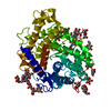
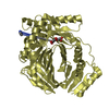
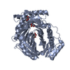
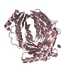
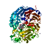
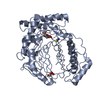
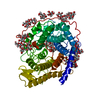
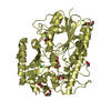
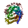
| Title | REFINED CRYSTAL STRUCTURES OF GLUCOAMYLASE FROM ASPERGILLUS AWAMORI VAR. X100 | |||||||||
 Components Components | GLUCOAMYLASE-471 | |||||||||
 Keywords Keywords | HYDROLASE | |||||||||
| Function / homology |  Function and homology information Function and homology informationglucan 1,4-alpha-glucosidase / glucan 1,4-alpha-glucosidase activity / starch binding / fungal-type vacuole / polysaccharide catabolic process Similarity search - Function | |||||||||
| Biological species |  | |||||||||
| Method |  X-RAY DIFFRACTION / Resolution: 2.4 Å X-RAY DIFFRACTION / Resolution: 2.4 Å | |||||||||
 Authors Authors | Aleshin, A.E. / Hoffman, C. / Firsov, L.M. / Honzatko, R.B. | |||||||||
 Citation Citation | Journal: J.Mol.Biol. / Year: 1994 Title: Refined crystal structures of glucoamylase from Aspergillus awamori var. X100. Authors: Aleshin, A.E. / Hoffman, C. / Firsov, L.M. / Honzatko, R.B. #1: Journal: To be PublishedTitle: Refined Structure for the Complex of Acarbose with Glucoamylase from Aspergillus Awamori Var. X100 to 2.4A Resolution Authors: Aleshin, A.E. / Firsov, L.M. / Honzatko, R.B. #2: Journal: Biochemistry / Year: 1993Title: Refined Structure of the Complex of 1-Deoxynojirimycin with Glucoamylase from Aspergillus Awamori Var. X100 to 2.4 Angstroms Resolution Authors: Harris, E.M.S. / Aleshin, A.E. / Firsov, L.M. / Honzatko, R.B. #3: Journal: J.Biol.Chem. / Year: 1992Title: Crystal Structure of Glucoamylase from Aspergillus Awamori Var. X100 Authors: Aleshin, A.E. / Golubev, A. / Firsov, L.M. / Honzatko, R.B. | |||||||||
| History |
|
- Structure visualization
| Structure viewer | Molecule: MolmilJmol/JSmol |
|---|
- Downloads & links
-Download
| PDBx/mmCIF format | 1glm.cif.gz | 122.8 KB | Display | PDBx/mmCIF format |
|---|---|---|---|---|
| PDB format | pdb1glm.ent.gz | 93 KB | Display | PDB format |
| PDBx/mmJSON format | 1glm.json.gz | Tree view | PDBx/mmJSON format | |
| Others |  Other downloads Other downloads |
-Validation report
| Arichive directory | https://data.pdbj.org/pub/pdb/validation_reports/gl/1glmftp://data.pdbj.org/pub/pdb/validation_reports/gl/1glm | HTTPS FTP |
|---|
-Related structure data


-Links
 PDBj
PDBj


- Assembly
| Deposited unit | 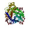
| ||||||||
|---|---|---|---|---|---|---|---|---|---|
| 1 |
| ||||||||
| Unit cell |
| ||||||||
| Atom site foot note | 1: GLY 23 - ALA 24 OMEGA = 1.53 PEPTIDE BOND DEVIATES SIGNIFICANTLY FROM TRANS CONFORMATION 2: CIS PROLINE - PRO 46 / 3: CIS PROLINE - PRO 123 4: RESIDUES ASN 171 AND ASN 395 ARE SITES OF N-GLYCOSYLATION. 5: RESIDUES SER 443, SER 444, THR 452, SER 453, SER 455, THR 457, SER 459, SER 460, THR 462, AND THR 464 ARE SITES OF O-GLYCOSYLATION. |
-Components
| #1: Protein | Mass: 50450.730 Da / Num. of mol.: 1 Source method: isolated from a genetically manipulated source Source: (gene. exp.) | ||||||
|---|---|---|---|---|---|---|---|
| #2: Polysaccharide | alpha-D-mannopyranose-(1-2)-alpha-D-mannopyranose-(1-3)-alpha-D-mannopyranose-(1-4)-2-acetamido-2- ...alpha-D-mannopyranose-(1-2)-alpha-D-mannopyranose-(1-3)-alpha-D-mannopyranose-(1-4)-2-acetamido-2-deoxy-beta-D-glucopyranose-(1-4)-2-acetamido-2-deoxy-beta-D-glucopyranose Source method: isolated from a genetically manipulated source | ||||||
| #3: Polysaccharide | alpha-D-mannopyranose-(1-2)-alpha-D-mannopyranose-(1-2)-alpha-D-mannopyranose-(1-3)-[alpha-D- ...alpha-D-mannopyranose-(1-2)-alpha-D-mannopyranose-(1-2)-alpha-D-mannopyranose-(1-3)-[alpha-D-mannopyranose-(1-3)-alpha-D-mannopyranose-(1-6)]alpha-D-mannopyranose-(1-4)-2-acetamido-2-deoxy-beta-D-glucopyranose-(1-4)-2-acetamido-2-deoxy-beta-D-glucopyranose Source method: isolated from a genetically manipulated source | ||||||
| #4: Sugar | ChemComp-MAN /   Type: D-saccharide, alpha linking / Mass: 180.156 Da / Num. of mol.: 10 Type: D-saccharide, alpha linking / Mass: 180.156 Da / Num. of mol.: 10Source method: isolated from a genetically manipulated source Formula: C6H12O6 #5: Water | ChemComp-HOH / |  Mass: 18.015 Da / Num. of mol.: 561 / Source method: isolated from a natural source / Formula: H2O Mass: 18.015 Da / Num. of mol.: 561 / Source method: isolated from a natural source / Formula: H2OHas protein modification | Y | Sequence details | RESIDUE NUMBER 102 IS SKIPPED SO THAT THE NUMBERING OF THE RESIDUES IS CONSISTENT WITH GLUCOAMYLASE ...RESIDUE NUMBER 102 IS SKIPPED SO THAT THE NUMBERING OF THE RESIDUES IS CONSISTENT | |
-Experimental details
-Experiment
| Experiment | Method: X-RAY DIFFRACTION |
|---|
- Sample preparation
| Crystal | Density Matthews: 2.93 Å3/Da / Density % sol: 58.06 % | ||||||||||||||||||||||||||||||
|---|---|---|---|---|---|---|---|---|---|---|---|---|---|---|---|---|---|---|---|---|---|---|---|---|---|---|---|---|---|---|---|
| Crystal grow | *PLUS pH: 5.95 / Method: vapor diffusion | ||||||||||||||||||||||||||||||
| Components of the solutions | *PLUS
|
-Data collection
| Radiation | Scattering type: x-ray |
|---|---|
| Radiation wavelength | Relative weight: 1 |
| Reflection | *PLUS Highest resolution: 2.4 Å / Num. all: 24005 / Num. obs: 20905 / Num. measured all: 57478 / Rmerge(I) obs: 0.035 |
- Processing
| Software | Name: PROLSQ / Classification: refinement | ||||||||||||||||||||||||||||||||||||||||||||||||||||||||||||||||||||||||||||||||||||
|---|---|---|---|---|---|---|---|---|---|---|---|---|---|---|---|---|---|---|---|---|---|---|---|---|---|---|---|---|---|---|---|---|---|---|---|---|---|---|---|---|---|---|---|---|---|---|---|---|---|---|---|---|---|---|---|---|---|---|---|---|---|---|---|---|---|---|---|---|---|---|---|---|---|---|---|---|---|---|---|---|---|---|---|---|---|
| Refinement | Resolution: 2.4→10 Å / σ(F): 1 /
| ||||||||||||||||||||||||||||||||||||||||||||||||||||||||||||||||||||||||||||||||||||
| Refinement step | Cycle: LAST / Resolution: 2.4→10 Å
| ||||||||||||||||||||||||||||||||||||||||||||||||||||||||||||||||||||||||||||||||||||
| Refine LS restraints |
| ||||||||||||||||||||||||||||||||||||||||||||||||||||||||||||||||||||||||||||||||||||
| Software | *PLUS Name: PROLSQ / Classification: refinement | ||||||||||||||||||||||||||||||||||||||||||||||||||||||||||||||||||||||||||||||||||||
| Refinement | *PLUS Rfactor obs: 0.122 | ||||||||||||||||||||||||||||||||||||||||||||||||||||||||||||||||||||||||||||||||||||
| Solvent computation | *PLUS | ||||||||||||||||||||||||||||||||||||||||||||||||||||||||||||||||||||||||||||||||||||
| Displacement parameters | *PLUS |
