 Movie
Movie Controller


+ Open data

- Basic information
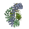
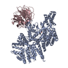
| Entry | Database: PDB / ID: 1gkr | ||||||
|---|---|---|---|---|---|---|---|
| Title | L-Hydantoinase (Dihydropyrimidinase) from Arthrobacter aurescens | ||||||
 Components Components | NON-ATP DEPENDENT L-SELECTIVE HYDANTOINASE | ||||||
 Keywords Keywords | HYDROLASE / HYDANTOINASE / DIHYDROPYRIMIDINASE / CYCLIC AMIDASE | ||||||
| Function / homology |  Function and homology information Function and homology informationHydrolases; Acting on carbon-nitrogen bonds, other than peptide bonds; In cyclic amides / allantoin catabolic process / allantoinase activity / purine nucleobase catabolic process / cobalt ion binding / zinc ion binding / cytoplasm Similarity search - Function | ||||||
| Biological species |  ARTHROBACTER AURESCENS (bacteria) ARTHROBACTER AURESCENS (bacteria) | ||||||
| Method |  X-RAY DIFFRACTION / MOLECULAR REPLACEMENT / Resolution: 2.6 Å X-RAY DIFFRACTION / MOLECULAR REPLACEMENT / Resolution: 2.6 Å | ||||||
 Authors Authors | Abendroth, J. / Niefind, K. / Schomburg, D. | ||||||
 Citation Citation | Journal: Biochemistry / Year: 2002 Title: The structure of L-hydantoinase from Arthobacter aurescens leads to an understanding of dihydropyrimidinase substrate and enantio specificity. Authors: Abendroth, J. / Niefind, K. / May, O. / Siemann, M. / Syldatk, C. / Schomburg, D. #1: Journal: Acta Crystallogr.,Sect.D / Year: 1996 Title: Crystallization and Preliminary X-Ray Analysis of a Hydantoinase from Arthrobacter Aurescens Dsm 3745. Authors: May, O. / Siemann, M. / Syldatk, C. / Niefind, K. / Schomburg, D. | ||||||
| History |
| ||||||
| Remark 700 | SHEET THE SHEET STRUCTURE OF THIS MOLECULE IS BIFURCATED. IN ORDER TO REPRESENT THIS FEATURE IN ... SHEET THE SHEET STRUCTURE OF THIS MOLECULE IS BIFURCATED. IN ORDER TO REPRESENT THIS FEATURE IN THE SHEET RECORDS BELOW, TWO SHEETS ARE DEFINED. |
- Structure visualization
| Structure viewer | Molecule: MolmilJmol/JSmol |
|---|
- Downloads & links
-Download
| PDBx/mmCIF format | 1gkr.cif.gz | 351 KB | Display | PDBx/mmCIF format |
|---|---|---|---|---|
| PDB format | pdb1gkr.ent.gz | 287.9 KB | Display | PDB format |
| PDBx/mmJSON format | 1gkr.json.gz | Tree view | PDBx/mmJSON format | |
| Others |  Other downloads Other downloads |
-Validation report
| Arichive directory | https://data.pdbj.org/pub/pdb/validation_reports/gk/1gkrftp://data.pdbj.org/pub/pdb/validation_reports/gk/1gkr | HTTPS FTP |
|---|
-Related structure data
| Related structure data |  1gkpS S: Starting model for refinement |
|---|---|
| Similar structure data |


-Links
 PDBj
PDBj

- Assembly
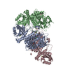
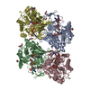
| Deposited unit | 
| ||||||||||||||||
|---|---|---|---|---|---|---|---|---|---|---|---|---|---|---|---|---|---|
| 1 |
| ||||||||||||||||
| Unit cell |
| ||||||||||||||||
| Noncrystallographic symmetry (NCS) | NCS oper:
|
-Components
| #1: Protein | Mass: 49693.445 Da / Num. of mol.: 4 / Source method: isolated from a natural source / Details: DSM 3745 / Source: (natural) ARTHROBACTER AURESCENS (bacteria) / References: UniProt: P81006, dihydropyrimidinase#2: Chemical | ChemComp-ZN /   Mass: 65.409 Da / Num. of mol.: 8 / Source method: obtained synthetically / Formula: Zn Mass: 65.409 Da / Num. of mol.: 8 / Source method: obtained synthetically / Formula: Zn#3: Water | ChemComp-HOH / |  Mass: 18.015 Da / Num. of mol.: 508 / Source method: isolated from a natural source / Formula: H2O Mass: 18.015 Da / Num. of mol.: 508 / Source method: isolated from a natural source / Formula: H2O |
|---|
-Experimental details
-Experiment
| Experiment | Method: X-RAY DIFFRACTION / Number of used crystals: 2 |
|---|
- Sample preparation
| Crystal | Density Matthews: 1.2 Å3/Da / Density % sol: 63 % | |||||||||||||||||||||||||||||||||||||||||||||||||
|---|---|---|---|---|---|---|---|---|---|---|---|---|---|---|---|---|---|---|---|---|---|---|---|---|---|---|---|---|---|---|---|---|---|---|---|---|---|---|---|---|---|---|---|---|---|---|---|---|---|---|
| Crystal grow | pH: 8.5 Details: 16-18% (W/V) PEG 8000, 250 MM LITHIUM SULPHATE, 100 MM MES/TRIS PH 8.5 | |||||||||||||||||||||||||||||||||||||||||||||||||
| Crystal grow | *PLUS Temperature: 292 K / pH: 8 / Method: vapor diffusion, sitting dropDetails: May, O., (1996) Acta Crystallogr.,Sect.D, D52, 1209. | |||||||||||||||||||||||||||||||||||||||||||||||||
| Components of the solutions | *PLUS
|
-Data collection
| Diffraction | Mean temperature: 283 K |
|---|---|
| Diffraction source | Source: ROTATING ANODE / Type: RIGAKU / Wavelength: 1.5418 |
| Detector | Type: SIEMENS / Detector: AREA DETECTOR / Date: Mar 20, 1996 |
| Radiation | Monochromator: GRAPHITE CRYSTAL / Protocol: SINGLE WAVELENGTH / Monochromatic (M) / Laue (L): M / Scattering type: x-ray |
| Radiation wavelength | Wavelength: 1.5418 Å / Relative weight: 1 |
| Reflection | Resolution: 2.6→20 Å / Num. obs: 61763 / % possible obs: 86.6 % / Redundancy: 2 % / Rmerge(I) obs: 0.091 / Net I/σ(I): 8.5 |
| Reflection shell | Resolution: 2.6→2.74 Å / Redundancy: 1.3 % / Rmerge(I) obs: 0.148 / Mean I/σ(I) obs: 2.5 / % possible all: 41.2 |
| Reflection | *PLUS Highest resolution: 2.6 Å / Lowest resolution: 20 Å / Num. measured all: 124261 |
| Reflection shell | *PLUS % possible obs: 41.2 % |
- Processing
| Software |
| ||||||||||||||||||||||||||||||||||||||||||||||||||||||||||||
|---|---|---|---|---|---|---|---|---|---|---|---|---|---|---|---|---|---|---|---|---|---|---|---|---|---|---|---|---|---|---|---|---|---|---|---|---|---|---|---|---|---|---|---|---|---|---|---|---|---|---|---|---|---|---|---|---|---|---|---|---|---|
| Refinement | Method to determine structure: MOLECULAR REPLACEMENT Starting model: HOMOLOGY MODEL FROM PDB ENTRY 1GKP Resolution: 2.6→30 Å / Rfactor Rfree error: 0.0043 / Data cutoff high absF: 10000 / Cross valid method: THROUGHOUT / σ(F): 0 Details: THE STRUCTURE HAS BEEN REFINED WITH ONLY ONE CHAIN USING THE NCS MATRICES GIVEN BELOW
| ||||||||||||||||||||||||||||||||||||||||||||||||||||||||||||
| Solvent computation | Bsol: 30.7 Å2 / ksol: 0.295 e/Å3 | ||||||||||||||||||||||||||||||||||||||||||||||||||||||||||||
| Displacement parameters | Biso mean: 23.6 Å2
| ||||||||||||||||||||||||||||||||||||||||||||||||||||||||||||
| Refine analyze |
| ||||||||||||||||||||||||||||||||||||||||||||||||||||||||||||
| Refinement step | Cycle: LAST / Resolution: 2.6→30 Å
| ||||||||||||||||||||||||||||||||||||||||||||||||||||||||||||
| Refine LS restraints |
| ||||||||||||||||||||||||||||||||||||||||||||||||||||||||||||
| Refine LS restraints NCS | NCS model details: CONSTRAINTS | ||||||||||||||||||||||||||||||||||||||||||||||||||||||||||||
| LS refinement shell | Resolution: 2.6→2.69 Å / Rfactor Rfree error: 0.034 / Total num. of bins used: 10
| ||||||||||||||||||||||||||||||||||||||||||||||||||||||||||||
| Refinement | *PLUS Lowest resolution: 30 Å / Rfactor Rfree: 0.243 | ||||||||||||||||||||||||||||||||||||||||||||||||||||||||||||
| Solvent computation | *PLUS | ||||||||||||||||||||||||||||||||||||||||||||||||||||||||||||
| Displacement parameters | *PLUS | ||||||||||||||||||||||||||||||||||||||||||||||||||||||||||||
| Refine LS restraints | *PLUS
|
