 Movie
Movie Controller

[English] 日本語
 Yorodumi
- PDB-1gej: STRUCTURAL CHARACTERIZATION OF N-BUTYL-ISOCYANIDE COMPLEXES OF CY... -
+ Open data

- Basic information
| Entry | Database: PDB / ID: 1gej | ||||||
|---|---|---|---|---|---|---|---|
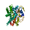
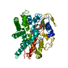
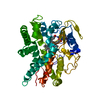
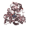
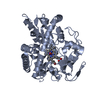
| Title | STRUCTURAL CHARACTERIZATION OF N-BUTYL-ISOCYANIDE COMPLEXES OF CYTOCHROMES P450NOR AND P450CAM | ||||||
 Components Components | CYTOCHROME P450 55A1 | ||||||
 Keywords Keywords | OXIDOREDUCTASE / Cytocrome P450nor (Fe-III) / Isocyanide Complexes form | ||||||
| Function / homology |  Function and homology information Function and homology informationnitric oxide reductase [NAD(P)+, nitrous oxide-forming] / nitric oxide reductase (NAD(P)H) activity / cholest-4-en-3-one 26-monooxygenase activity / steroid hydroxylase activity / cholesterol catabolic process / iron ion binding / heme binding Similarity search - Function | ||||||
| Biological species |   Fusarium oxysporum (fungus) Fusarium oxysporum (fungus) | ||||||
| Method |  X-RAY DIFFRACTION / SYNCHROTRON / MOLECULAR REPLACEMENT / Resolution: 1.5 Å X-RAY DIFFRACTION / SYNCHROTRON / MOLECULAR REPLACEMENT / Resolution: 1.5 Å | ||||||
 Authors Authors | lee, D.-S. / Park, S.-Y. / Yamane, K. / Shiro, Y. | ||||||
 Citation Citation | Journal: Biochemistry / Year: 2001 Title: Structural characterization of n-butyl-isocyanide complexes of cytochromes P450nor and P450cam. Authors: Lee, D.S. / Park, S.Y. / Yamane, K. / Obayashi, E. / Hori, H. / Shiro, Y. #1: Journal: Nat.Struct.Biol. / Year: 1997Title: Crystal structure of nitric oxide reductase from denitrifing fungus Fusarium oxysporum Authors: Park, S.-Y. / Shimizu, H. / Adachi, S. / Nakagawa, A. / Tanaka, I. / Nakahara, K. / Shoun, H. / Obayashi, E. / Nakamura, H. / Iizuka, T. / Shiro, Y. | ||||||
| History |
|
- Structure visualization
| Structure viewer | Molecule: MolmilJmol/JSmol |
|---|
- Downloads & links
-Download
| PDBx/mmCIF format | 1gej.cif.gz | 98.8 KB | Display | PDBx/mmCIF format |
|---|---|---|---|---|
| PDB format | pdb1gej.ent.gz | 72.9 KB | Display | PDB format |
| PDBx/mmJSON format | 1gej.json.gz | Tree view | PDBx/mmJSON format | |
| Others |  Other downloads Other downloads |
-Validation report
| Summary document | 1gej_validation.pdf.gz | 478.2 KB | Display | wwPDB validaton report |
|---|---|---|---|---|
| Full document | 1gej_full_validation.pdf.gz | 479.9 KB | Display | |
| Data in XML | 1gej_validation.xml.gz | 9.3 KB | Display | |
| Data in CIF | 1gej_validation.cif.gz | 15.6 KB | Display | |
| Arichive directory | https://data.pdbj.org/pub/pdb/validation_reports/ge/1gejftp://data.pdbj.org/pub/pdb/validation_reports/ge/1gej | HTTPS FTP |
-Related structure data
| Related structure data |  1geiC 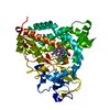 1gekC 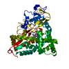 1gemC  1romS C: citing same article ( S: Starting model for refinement |
|---|---|
| Similar structure data |


-Links
 PDBj
PDBj
- Assembly
| Deposited unit | 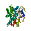
| ||||||||
|---|---|---|---|---|---|---|---|---|---|
| 1 |
| ||||||||
| Unit cell |
|
-Components
| #1: Protein | Mass: 44420.691 Da / Num. of mol.: 1 Source method: isolated from a genetically manipulated source Source: (gene. exp.) Fusarium oxysporum (fungus) / Plasmid: PRSET-C / Production host:  References: UniProt: P23295, Oxidoreductases; Acting on paired donors, with incorporation or reduction of molecular oxygen |
|---|---|
| #2: Chemical | ChemComp-HEM /   Mass: 616.487 Da / Num. of mol.: 1 / Source method: obtained synthetically / Formula: C34H32FeN4O4 Mass: 616.487 Da / Num. of mol.: 1 / Source method: obtained synthetically / Formula: C34H32FeN4O4 |
| #3: Chemical | ChemComp-NBN /   Mass: 83.132 Da / Num. of mol.: 1 / Source method: obtained synthetically / Formula: C5H9N Mass: 83.132 Da / Num. of mol.: 1 / Source method: obtained synthetically / Formula: C5H9N |
| #4: Water | ChemComp-HOH /  Mass: 18.015 Da / Num. of mol.: 303 / Source method: isolated from a natural source / Formula: H2O Mass: 18.015 Da / Num. of mol.: 303 / Source method: isolated from a natural source / Formula: H2O |
-Experimental details
-Experiment
| Experiment | Method: X-RAY DIFFRACTION / Number of used crystals: 1 |
|---|
- Sample preparation
| Crystal | Density Matthews: 2.16 Å3/Da / Density % sol: 43.01 % | ||||||||||||||||||||||||||||||||||||||||
|---|---|---|---|---|---|---|---|---|---|---|---|---|---|---|---|---|---|---|---|---|---|---|---|---|---|---|---|---|---|---|---|---|---|---|---|---|---|---|---|---|---|
| Crystal grow | Temperature: 293 K / Method: vapor diffusion, sitting drop / pH: 6.5 Details: PEG4000, pH 6.5, VAPOR DIFFUSION, SITTING DROP, temperature 293K | ||||||||||||||||||||||||||||||||||||||||
| Crystal grow | *PLUS Temperature: 4 ℃ / pH: 7.2 / Details: Park, S-Y., (1997) FEBS. Lett., 412, 346. | ||||||||||||||||||||||||||||||||||||||||
| Components of the solutions | *PLUS
|
-Data collection
| Diffraction | Mean temperature: 100 K |
|---|---|
| Diffraction source | Source: SYNCHROTRON / Site: SPring-8  / Beamline: BL44B2 / Wavelength: 0.7 Å / Beamline: BL44B2 / Wavelength: 0.7 Å |
| Detector | Type: MAR CCD 165 mm / Detector: CCD / Date: Feb 20, 2000 / Details: mirrors |
| Radiation | Monochromator: 0.7 / Protocol: SINGLE WAVELENGTH / Monochromatic (M) / Laue (L): M / Scattering type: x-ray |
| Radiation wavelength | Wavelength: 0.7 Å / Relative weight: 1 |
| Reflection | Resolution: 1.5→25 Å / Num. all: 356663 / Num. obs: 57289 / % possible obs: 92.4 % / Observed criterion σ(F): 0 / Observed criterion σ(I): 0 / Redundancy: 6.2 % / Biso Wilson estimate: 15.4 Å2 / Rmerge(I) obs: 0.056 / Net I/σ(I): 9.8 |
| Reflection shell | Resolution: 1.5→1.58 Å / Redundancy: 4.6 % / Rmerge(I) obs: 0.278 / Mean I/σ(I) obs: 2.8 / Num. unique all: 5725 / % possible all: 65.3 |
| Reflection shell | *PLUS % possible obs: 65.3 % |
- Processing
| Software |
| |||||||||||||||||||||||||
|---|---|---|---|---|---|---|---|---|---|---|---|---|---|---|---|---|---|---|---|---|---|---|---|---|---|---|
| Refinement | Method to determine structure: MOLECULAR REPLACEMENT Starting model: PDB ENTRY 1ROM Resolution: 1.5→25 Å / Isotropic thermal model: Isotropic / Cross valid method: THROUGHOUT / σ(F): 0 / σ(I): 0 / Stereochemistry target values: Engh & Huber
| |||||||||||||||||||||||||
| Displacement parameters | Biso mean: 15.4 Å2 | |||||||||||||||||||||||||
| Refine analyze |
| |||||||||||||||||||||||||
| Refinement step | Cycle: LAST / Resolution: 1.5→25 Å
| |||||||||||||||||||||||||
| Refine LS restraints |
| |||||||||||||||||||||||||
| LS refinement shell | Resolution: 1.5→1.59 Å / Rfactor Rfree error: 0.015
| |||||||||||||||||||||||||
| Software | *PLUS Name: X-PLOR / Version: 3.851 / Classification: refinement | |||||||||||||||||||||||||
| Refine LS restraints | *PLUS
|
