 Movie
Movie Controller

[English] 日本語
 Yorodumi
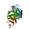
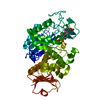
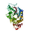
- PDB-1gcy: HIGH RESOLUTION CRYSTAL STRUCTURE OF MALTOTETRAOSE-FORMING EXO-AMYLASE -
+ Open data

- Basic information
| Entry | Database: PDB / ID: 1gcy | ||||||
|---|---|---|---|---|---|---|---|
| Title | HIGH RESOLUTION CRYSTAL STRUCTURE OF MALTOTETRAOSE-FORMING EXO-AMYLASE | ||||||
 Components Components | GLUCAN 1,4-ALPHA-MALTOTETRAHYDROLASE | ||||||
 Keywords Keywords | HYDROLASE / BETA-ALPHA-BARREL / BETA SHEET | ||||||
| Function / homology |  Function and homology information Function and homology informationglucan 1,4-alpha-maltotetraohydrolase / glucan 1,4-alpha-maltotetraohydrolase activity / starch catabolic process / starch binding / alpha-amylase activity / extracellular region / metal ion binding Similarity search - Function | ||||||
| Biological species |  Pseudomonas stutzeri (bacteria) Pseudomonas stutzeri (bacteria) | ||||||
| Method |  X-RAY DIFFRACTION / SYNCHROTRON / MOLECULAR REPLACEMENT / Resolution: 1.6 Å X-RAY DIFFRACTION / SYNCHROTRON / MOLECULAR REPLACEMENT / Resolution: 1.6 Å | ||||||
 Authors Authors | Mezaki, Y. / Katsuya, Y. / Kubota, M. / Matsuura, Y. | ||||||
 Citation Citation | Journal: Biosci.Biotechnol.Biochem. / Year: 2001 Title: Crystallization and structural analysis of intact maltotetraose-forming exo-amylase from Pseudomonas stutzeri. Authors: Mezaki, Y. / Katsuya, Y. / Kubota, M. / Matsuura, Y. #1: Journal: J.Mol.Biol. / Year: 1997Title: Crystal Structure of a Maltotetraose-forming Exo-amylase from Pseudomonas stutzeri Authors: Morishita, Y. / Hasegawa, K. / Matsuura, Y. / Katsube, Y. / Kubota, M. / Sakai, S. #2: Journal: J.Mol.Biol. / Year: 1997Title: Crystal Structure of a Mutant Maltotetraose-forming Exo-amylase Cocrystallized with Maltopentaose Authors: Yoshioka, Y. / Hasegawa, K. / Matsuura, Y. / Katsube, Y. / Kubota, M. | ||||||
| History |
|
- Structure visualization
| Structure viewer | Molecule: MolmilJmol/JSmol |
|---|
- Downloads & links
-Download
| PDBx/mmCIF format | 1gcy.cif.gz | 101 KB | Display | PDBx/mmCIF format |
|---|---|---|---|---|
| PDB format | pdb1gcy.ent.gz | 75.1 KB | Display | PDB format |
| PDBx/mmJSON format | 1gcy.json.gz | Tree view | PDBx/mmJSON format | |
| Others |  Other downloads Other downloads |
-Validation report
| Arichive directory | https://data.pdbj.org/pub/pdb/validation_reports/gc/1gcyftp://data.pdbj.org/pub/pdb/validation_reports/gc/1gcy | HTTPS FTP |
|---|
-Related structure data
| Related structure data | 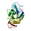 2amgS S: Starting model for refinement |
|---|---|
| Similar structure data |


-Links
 PDBj
PDBj

- Assembly
| Deposited unit | 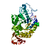
| ||||||||
|---|---|---|---|---|---|---|---|---|---|
| 1 |
| ||||||||
| Unit cell |
|
-Components
| #1: Protein | Mass: 57769.004 Da / Num. of mol.: 1 Source method: isolated from a genetically manipulated source Source: (gene. exp.) Pseudomonas stutzeri (bacteria) / Strain: MO-19 / Production host: References: UniProt: P13507, glucan 1,4-alpha-maltotetraohydrolase | ||||||
|---|---|---|---|---|---|---|---|
| #2: Chemical |   Mass: 40.078 Da / Num. of mol.: 2 / Source method: obtained synthetically / Formula: Ca Mass: 40.078 Da / Num. of mol.: 2 / Source method: obtained synthetically / Formula: Ca#3: Water | ChemComp-HOH / |  Mass: 18.015 Da / Num. of mol.: 222 / Source method: isolated from a natural source / Formula: H2O Mass: 18.015 Da / Num. of mol.: 222 / Source method: isolated from a natural source / Formula: H2OCompound details | The C-terminal raw-starch binding domain of about a hundred amino acids is disordered in the crystal. | Has protein modification | Y | |
-Experimental details
-Experiment
| Experiment | Method: X-RAY DIFFRACTION / Number of used crystals: 1 |
|---|
- Sample preparation
| Crystal | Density Matthews: 2.39 Å3/Da / Density % sol: 48.52 % | ||||||||||||||||||||
|---|---|---|---|---|---|---|---|---|---|---|---|---|---|---|---|---|---|---|---|---|---|
| Crystal grow | Temperature: 277 K / Method: vapor diffusion, hanging drop / pH: 7.5 Details: ammonium sulfate, pH 7.5, VAPOR DIFFUSION, HANGING DROP, temperature 277K | ||||||||||||||||||||
| Crystal grow | *PLUS Temperature: 4 ℃ / Method: vapor diffusion | ||||||||||||||||||||
| Components of the solutions | *PLUS
|
-Data collection
| Diffraction | Mean temperature: 100 K |
|---|---|
| Diffraction source | Source: SYNCHROTRON / Site: SPring-8  / Beamline: BL24XU / Wavelength: 0.834 / Beamline: BL24XU / Wavelength: 0.834 |
| Detector | Type: RIGAKU RAXIS IV / Detector: IMAGE PLATE / Date: May 14, 1999 |
| Radiation | Protocol: SINGLE WAVELENGTH / Monochromatic (M) / Laue (L): M / Scattering type: x-ray |
| Radiation wavelength | Wavelength: 0.834 Å / Relative weight: 1 |
| Reflection | Highest resolution: 1.6 Å / Num. obs: 66492 / % possible obs: 89.6 % / Observed criterion σ(I): 1 / Redundancy: 3.3 % / Biso Wilson estimate: 19.4 Å2 / Rmerge(I) obs: 0.061 |
| Reflection | *PLUS Num. measured all: 222219 |
- Processing
| Software |
| ||||||||||||||||||||||||||||||||||||
|---|---|---|---|---|---|---|---|---|---|---|---|---|---|---|---|---|---|---|---|---|---|---|---|---|---|---|---|---|---|---|---|---|---|---|---|---|---|
| Refinement | Method to determine structure: MOLECULAR REPLACEMENT Starting model: 2AMG Resolution: 1.6→8 Å / Rfactor Rfree error: 0.005 / Data cutoff high absF: 1000000 / Data cutoff low absF: 0 / Isotropic thermal model: RESTRAINED / Cross valid method: THROUGHOUT / σ(F): 2
| ||||||||||||||||||||||||||||||||||||
| Displacement parameters | Biso mean: 19.4 Å2
| ||||||||||||||||||||||||||||||||||||
| Refine analyze |
| ||||||||||||||||||||||||||||||||||||
| Refinement step | Cycle: LAST / Resolution: 1.6→8 Å
| ||||||||||||||||||||||||||||||||||||
| Refine LS restraints |
| ||||||||||||||||||||||||||||||||||||
| LS refinement shell | Resolution: 1.6→1.7 Å / Rfactor Rfree error: 0.016 / Total num. of bins used: 6
| ||||||||||||||||||||||||||||||||||||
| Xplor file |
| ||||||||||||||||||||||||||||||||||||
| Software | *PLUS Name: X-PLOR / Version: 3.851 / Classification: refinement | ||||||||||||||||||||||||||||||||||||
| Refine LS restraints | *PLUS
|
