 Movie
Movie Controller


+ Open data

- Basic information
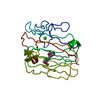
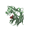
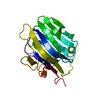
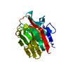
| Entry | Database: PDB / ID: 1gbg | ||||||
|---|---|---|---|---|---|---|---|
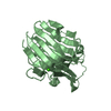
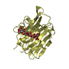
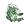
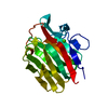
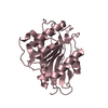
| Title | BACILLUS LICHENIFORMIS BETA-GLUCANASE | ||||||
 Components Components | (1,3-1,4)-BETA-D-GLUCAN 4 GLUCANOHYDROLASE | ||||||
 Keywords Keywords | HYDROLASE (GLUCANASE) | ||||||
| Function / homology |  Function and homology information Function and homology information | ||||||
| Biological species |  | ||||||
| Method |  X-RAY DIFFRACTION / Resolution: 1.8 Å X-RAY DIFFRACTION / Resolution: 1.8 Å | ||||||
 Authors Authors | Hahn, M. / Heinemann, U. | ||||||
 Citation Citation | Journal: FEBS Lett. / Year: 1995 Title: Crystal structure of Bacillus licheniformis 1,3-1,4-beta-D-glucan 4-glucanohydrolase at 1.8 A resolution. Authors: Hahn, M. / Pons, J. / Planas, A. / Querol, E. / Heinemann, U. #1: Journal: Proc.Natl.Acad.Sci.USA / Year: 1993Title: Molecular and Active-Site Structure of a Bacillus 1,3-1,4-Beta-Glucanase Authors: Keitel, T. / Simon, O. / Borriss, R. / Heinemann, U. #2: Journal: Eur.J.Biochem. / Year: 1991Title: Molecular Cloning, Expression and Nucleotide Sequence of the Endo-Beta-1,3-1,4-D-Glucanase Gene from Bacillus Licheniformis Authors: Lloberas, J. / Perez-Pons, J.A. / Querol, E. | ||||||
| History |
|
- Structure visualization
| Structure viewer | Molecule: MolmilJmol/JSmol |
|---|
- Downloads & links
-Download
| PDBx/mmCIF format | 1gbg.cif.gz | 59.3 KB | Display | PDBx/mmCIF format |
|---|---|---|---|---|
| PDB format | pdb1gbg.ent.gz | 42.3 KB | Display | PDB format |
| PDBx/mmJSON format | 1gbg.json.gz | Tree view | PDBx/mmJSON format | |
| Others |  Other downloads Other downloads |
-Validation report
| Arichive directory | https://data.pdbj.org/pub/pdb/validation_reports/gb/1gbgftp://data.pdbj.org/pub/pdb/validation_reports/gb/1gbg | HTTPS FTP |
|---|
-Related structure data
| Similar structure data |
|---|

-Links
 PDBj
PDBj
- Assembly
| Deposited unit | 
| ||||||||
|---|---|---|---|---|---|---|---|---|---|
| 1 |
| ||||||||
| Unit cell |
| ||||||||
| Atom site foot note | 1: CIS PROLINE - PRO 201 |
-Components
| #1: Protein | ( Mass: 24374.854 Da / Num. of mol.: 1 Source method: isolated from a genetically manipulated source Source: (gene. exp.) | ||
|---|---|---|---|
| #2: Chemical | ChemComp-CA /   Mass: 40.078 Da / Num. of mol.: 1 / Source method: obtained synthetically / Formula: Ca Mass: 40.078 Da / Num. of mol.: 1 / Source method: obtained synthetically / Formula: Ca | ||
| #3: Water | ChemComp-HOH /  Mass: 18.015 Da / Num. of mol.: 158 / Source method: isolated from a natural source / Formula: H2O Mass: 18.015 Da / Num. of mol.: 158 / Source method: isolated from a natural source / Formula: H2O | ||
| Compound details | BOND LENGTHS OF SIDE CHAINS OF GLU AND ASP FOLLOW THE TNT STANDARD FOR PROTONATED| Has protein modification | Y | |
-Experimental details
-Experiment
| Experiment | Method: X-RAY DIFFRACTION |
|---|
- Sample preparation
| Crystal | Density Matthews: 2.08 Å3/Da / Density % sol: 40.91 % | ||||||||||||||||||||||||||||||||||||
|---|---|---|---|---|---|---|---|---|---|---|---|---|---|---|---|---|---|---|---|---|---|---|---|---|---|---|---|---|---|---|---|---|---|---|---|---|---|
| Crystal | *PLUS Density % sol: 54 % | ||||||||||||||||||||||||||||||||||||
| Crystal grow | *PLUS pH: 7 / Method: vapor diffusion, hanging drop | ||||||||||||||||||||||||||||||||||||
| Components of the solutions | *PLUS
|
-Data collection
| Diffraction source | Wavelength: 1.5418 |
|---|---|
| Detector | Type: MARRESEARCH / Detector: IMAGE PLATE |
| Radiation | Monochromatic (M) / Laue (L): M / Scattering type: x-ray |
| Radiation wavelength | Wavelength: 1.5418 Å / Relative weight: 1 |
| Reflection | Redundancy: 1.9 % / Rmerge(I) obs: 0.082 |
| Reflection | *PLUS Highest resolution: 1.8 Å / Num. obs: 17238 / % possible obs: 94.6 % / Rmerge(I) obs: 0.082 |
- Processing
| Software |
| ||||||||||||||||||||||||||||||
|---|---|---|---|---|---|---|---|---|---|---|---|---|---|---|---|---|---|---|---|---|---|---|---|---|---|---|---|---|---|---|---|
| Refinement | Resolution: 1.8→6 Å / σ(F): 2 Details: ASN 28 IS IN CONTACT WITH A NEIGHBORING MOLECULE. THEREFORE ITS DIHEDRAL ANGLES LIE OUTSIDE THE EXPECTED RANGE.
| ||||||||||||||||||||||||||||||
| Refinement step | Cycle: LAST / Resolution: 1.8→6 Å
| ||||||||||||||||||||||||||||||
| Refine LS restraints |
| ||||||||||||||||||||||||||||||
| Software | *PLUS Name: TNT / Classification: refinement | ||||||||||||||||||||||||||||||
| Refinement | *PLUS Rfactor obs: 0.165 / Rfactor Rfree: 0.236 | ||||||||||||||||||||||||||||||
| Solvent computation | *PLUS | ||||||||||||||||||||||||||||||
| Displacement parameters | *PLUS | ||||||||||||||||||||||||||||||
| Refine LS restraints | *PLUS
|
