 Movie
Movie Controller


+ Open data

- Basic information
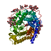
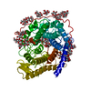
| Entry | Database: PDB / ID: 1gai | |||||||||
|---|---|---|---|---|---|---|---|---|---|---|
| Title | GLUCOAMYLASE-471 COMPLEXED WITH D-GLUCO-DIHYDROACARBOSE | |||||||||
 Components Components | GLUCOAMYLASE-471 | |||||||||
 Keywords Keywords | HYDROLASE / GLYCOSIDASE / POLYSACCHARIDE DEGRADATION / GLYCOPROTEIN | |||||||||
| Function / homology |  Function and homology information Function and homology informationglucan 1,4-alpha-glucosidase / glucan 1,4-alpha-glucosidase activity / starch binding / fungal-type vacuole / polysaccharide catabolic process Similarity search - Function | |||||||||
| Biological species |  | |||||||||
| Method |  X-RAY DIFFRACTION / MOLECULAR REPLACEMENT / Resolution: 1.7 Å X-RAY DIFFRACTION / MOLECULAR REPLACEMENT / Resolution: 1.7 Å | |||||||||
 Authors Authors | Aleshin, A.E. / Stoffer, B. / Firsov, L.M. / Svensson, B. / Honzatko, R.B. | |||||||||
 Citation Citation | Journal: Biochemistry / Year: 1996 Title: Crystallographic complexes of glucoamylase with maltooligosaccharide analogs: relationship of stereochemical distortions at the nonreducing end to the catalytic mechanism. Authors: Aleshin, A.E. / Stoffer, B. / Firsov, L.M. / Svensson, B. / Honzatko, R.B. #1: Journal: FEBS Lett. / Year: 1995Title: Refined Structure for the Complex of D-Gluco-Dihydroacarbose with Glucoamylase from Aspergillus Awamori Var. X100 to 2.2 A Resolution: Dual Conformations for Extended Inhibitors Bound to the ...Title: Refined Structure for the Complex of D-Gluco-Dihydroacarbose with Glucoamylase from Aspergillus Awamori Var. X100 to 2.2 A Resolution: Dual Conformations for Extended Inhibitors Bound to the Active Site of Glucoamylase Authors: Stoffer, B. / Aleshin, A.E. / Firsov, L.M. / Svensson, B. / Honzatko, R.B. #2: Journal: J.Mol.Biol. / Year: 1994Title: Refined Crystal Structures of Glucoamylase from Aspergillus Awamori Var. X100 Authors: Aleshin, A.E. / Hoffman, C. / Firsov, L.M. / Honzatko, R.B. #3: Journal: J.Biol.Chem. / Year: 1994Title: Refined Structure for the Complex of Acarbose with Glucoamylase from Aspergillus Awamori Var. X100 to 2.4-A Resolution Authors: Aleshin, A.E. / Firsov, L.M. / Honzatko, R.B. #4: Journal: Biochemistry / Year: 1993Title: Refined Structure for the Complex of 1-Deoxynojirimycin with Glucoamylase from Aspergillus Awamori Var. X100 to 2.4-A Resolution Authors: Harris, E.M. / Aleshin, A.E. / Firsov, L.M. / Honzatko, R.B. #5: Journal: J.Biol.Chem. / Year: 1992Title: Crystal Structure of Glucoamylase from Aspergillus Awamori Var. X100 to 2.2-A Resolution Authors: Aleshin, A. / Golubev, A. / Firsov, L.M. / Honzatko, R.B. | |||||||||
| History |
| |||||||||
| Remark 700 | SHEET MOST OF THE SHEETS FOR GLUCOAMYLASE-471 ARE HAIRPIN LOOPS THAT CONNECT HELICES. THESE LOOPS ...SHEET MOST OF THE SHEETS FOR GLUCOAMYLASE-471 ARE HAIRPIN LOOPS THAT CONNECT HELICES. THESE LOOPS HAVE TWO OR MORE H-BONDS BETWEEN THE ANTIPARALLEL STRANDS THAT COMPRISE THEM. IN ADDITION INDIVIDUAL LOOPS PACK TOGETHER, BUT THERE EXISTS GENERALLY ONLY ONE H-BOND BETWEEN A LOOP AND ITS NEIGHBOR. |
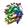
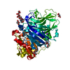
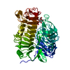
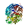
- Structure visualization
| Structure viewer | Molecule: MolmilJmol/JSmol |
|---|
- Downloads & links
-Download
| PDBx/mmCIF format | 1gai.cif.gz | 172.5 KB | Display | PDBx/mmCIF format |
|---|---|---|---|---|
| PDB format | pdb1gai.ent.gz | 141.7 KB | Display | PDB format |
| PDBx/mmJSON format | 1gai.json.gz | Tree view | PDBx/mmJSON format | |
| Others |  Other downloads Other downloads |
-Validation report
| Arichive directory | https://data.pdbj.org/pub/pdb/validation_reports/ga/1gaiftp://data.pdbj.org/pub/pdb/validation_reports/ga/1gai | HTTPS FTP |
|---|
-Related structure data


-Links
 PDBj
PDBj


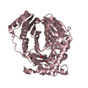
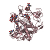
- Assembly
| Deposited unit | 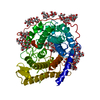
| ||||||||
|---|---|---|---|---|---|---|---|---|---|
| 1 |
| ||||||||
| Unit cell |
|
-Components
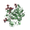
-Protein / Non-polymers , 2 types, 552 molecules A
| #1: Protein | Mass: 50608.883 Da / Num. of mol.: 1 / Fragment: RESIDUES 1-471 / Source method: isolated from a natural source / Details: COMPLEXED WITH D-GLUCO-DIHYDROACARBOSE / Source: (natural) References: UniProt: P22832, UniProt: P69327*PLUS, glucan 1,4-alpha-glucosidase |
|---|---|
| #6: Water | ChemComp-HOH / Mass: 18.015 Da / Num. of mol.: 551 / Source method: isolated from a natural source / Formula: H2O |
-Sugars , 4 types, 13 molecules 
| #2: Polysaccharide | alpha-D-mannopyranose-(1-2)-alpha-D-mannopyranose-(1-3)-beta-D-mannopyranose-(1-4)-2-acetamido-2- ...alpha-D-mannopyranose-(1-2)-alpha-D-mannopyranose-(1-3)-beta-D-mannopyranose-(1-4)-2-acetamido-2-deoxy-beta-D-glucopyranose-(1-4)-2-acetamido-2-deoxy-beta-D-glucopyranose Source method: isolated from a genetically manipulated source |
|---|---|
| #3: Polysaccharide | alpha-D-mannopyranose-(1-2)-alpha-D-mannopyranose-(1-2)-alpha-D-mannopyranose-(1-3)-[alpha-D- ...alpha-D-mannopyranose-(1-2)-alpha-D-mannopyranose-(1-2)-alpha-D-mannopyranose-(1-3)-[alpha-D-mannopyranose-(1-3)-[alpha-D-mannopyranose-(1-6)]alpha-D-mannopyranose-(1-6)]beta-D-mannopyranose-(1-4)-2-acetamido-2-deoxy-beta-D-glucopyranose-(1-4)-2-acetamido-2-deoxy-beta-D-glucopyranose Source method: isolated from a genetically manipulated source |
| #4: Polysaccharide | 4,6-dideoxy-4-{[(1S,2S,3S,4R,5R)-2,3,4-trihydroxy-5-(hydroxymethyl)cyclohexyl]amino}-alpha-D- ...4,6-dideoxy-4-{[(1S,2S,3S,4R,5R)-2,3,4-trihydroxy-5-(hydroxymethyl)cyclohexyl]amino}-alpha-D-glucopyranose-(1-4)-alpha-D-glucopyranose-(1-4)-alpha-D-glucopyranose / dihydro-alpha-acarbose  Type: oligosaccharide, Oligosaccharide / Class: Inhibitor / Mass: 647.622 Da / Num. of mol.: 1 Type: oligosaccharide, Oligosaccharide / Class: Inhibitor / Mass: 647.622 Da / Num. of mol.: 1Source method: isolated from a genetically manipulated source Details: oligosaccharide / References: dihydro-alpha-acarbose |
| #5: Sugar | ChemComp-MAN /  Type: D-saccharide, alpha linking / Mass: 180.156 Da / Num. of mol.: 10 Type: D-saccharide, alpha linking / Mass: 180.156 Da / Num. of mol.: 10Source method: isolated from a genetically manipulated source Formula: C6H12O6 |
-Details
| Compound details | GLUCOAMYLASE-471 IS A NATURAL PROTEOLYTIC FRAGMENT OF PARENT GLUCOAMYLASE, WHICH IS INITIALLY ...GLUCOAMYLA |
|---|---|
| Has protein modification | Y |
| Nonpolymer details | D-GLUCO-DIHYDROACARBOSE IS BOUND TO THE ACTIVE SITE OF THE ENZYME. D-GLUCO-DIHYDROACARBOSE IS A ...D-GLUCO-DIHYDROACA |
-Experimental details
-Experiment
| Experiment | Method: X-RAY DIFFRACTION / Number of used crystals: 1 |
|---|
- Sample preparation
| Crystal | Density Matthews: 2.8 Å3/Da / Density % sol: 56.1 % | ||||||||||||||||||||||||||||||
|---|---|---|---|---|---|---|---|---|---|---|---|---|---|---|---|---|---|---|---|---|---|---|---|---|---|---|---|---|---|---|---|
| Crystal grow | pH: 4 / Details: pH 4. | ||||||||||||||||||||||||||||||
| Crystal grow | *PLUS pH: 6.5 / Method: vapor diffusion, hanging drop / Details: Golubev, A. M., (1992) J. Mol. Biol., 226, 271. | ||||||||||||||||||||||||||||||
| Components of the solutions | *PLUS
|
-Data collection
| Diffraction | Mean temperature: 293 K |
|---|---|
| Diffraction source | Source: ROTATING ANODE / Type: SIEMENS / Wavelength: 1.5418 |
| Detector | Type: SIEMENS / Detector: AREA DETECTOR / Date: Oct 20, 1993 |
| Radiation | Monochromator: GRAPHITE(002) / Monochromatic (M) / Laue (L): M / Scattering type: x-ray |
| Radiation wavelength | Wavelength: 1.5418 Å / Relative weight: 1 |
| Reflection | Resolution: 1.69→30 Å / Num. obs: 64147 / % possible obs: 96 % / Observed criterion σ(I): 4 / Redundancy: 4.66 % / Rmerge(I) obs: 0.055 / Net I/σ(I): 36.16 |
| Reflection shell | Resolution: 1.69→1.8 Å / Redundancy: 1.9 % / Rmerge(I) obs: 0.257 / Mean I/σ(I) obs: 3.3 / % possible all: 75.5 |
| Reflection | *PLUS Num. obs: 64713 / Num. measured all: 298712 |
| Reflection shell | *PLUS Highest resolution: 1.7 Å / % possible obs: 75 % |
- Processing
| Software |
| ||||||||||||||||||||||||||||||||||||||||||||||||||||||||||||
|---|---|---|---|---|---|---|---|---|---|---|---|---|---|---|---|---|---|---|---|---|---|---|---|---|---|---|---|---|---|---|---|---|---|---|---|---|---|---|---|---|---|---|---|---|---|---|---|---|---|---|---|---|---|---|---|---|---|---|---|---|---|
| Refinement | Method to determine structure: MOLECULAR REPLACEMENT Starting model: NATIVE GLOCOAMYLASE Resolution: 1.7→10 Å / σ(F): 1
| ||||||||||||||||||||||||||||||||||||||||||||||||||||||||||||
| Displacement parameters | Biso mean: 14.7 Å2 | ||||||||||||||||||||||||||||||||||||||||||||||||||||||||||||
| Refine analyze | Luzzati coordinate error obs: 0.15 Å | ||||||||||||||||||||||||||||||||||||||||||||||||||||||||||||
| Refinement step | Cycle: LAST / Resolution: 1.7→10 Å
| ||||||||||||||||||||||||||||||||||||||||||||||||||||||||||||
| Refine LS restraints |
| ||||||||||||||||||||||||||||||||||||||||||||||||||||||||||||
| Xplor file |
| ||||||||||||||||||||||||||||||||||||||||||||||||||||||||||||
| Software | *PLUS Name: X-PLOR / Version: 3.1 / Classification: refinement | ||||||||||||||||||||||||||||||||||||||||||||||||||||||||||||
| Refine LS restraints | *PLUS
|
