 Movie
Movie Controller


+ Open data

- Basic information
| Entry | Database: PDB / ID: 1g9t | ||||||
|---|---|---|---|---|---|---|---|
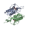
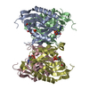
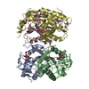
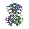
| Title | CRYSTAL STRUCTURE OF E.COLI HPRT-GMP COMPLEX | ||||||
 Components Components | HYPOXANTHINE PHOSPHORIBOSYLTRANSFERASE | ||||||
 Keywords Keywords | TRANSFERASE / phosphoribosyltransferases / purine salvage / protein chemistry / enzymology | ||||||
| Function / homology |  Function and homology information Function and homology informationhypoxanthine phosphoribosyltransferase / guanine phosphoribosyltransferase activity / guanine salvage / hypoxanthine metabolic process / hypoxanthine phosphoribosyltransferase activity / GMP salvage / IMP salvage / purine ribonucleoside salvage / guanosine tetraphosphate binding / protein homotetramerization ...hypoxanthine phosphoribosyltransferase / guanine phosphoribosyltransferase activity / guanine salvage / hypoxanthine metabolic process / hypoxanthine phosphoribosyltransferase activity / GMP salvage / IMP salvage / purine ribonucleoside salvage / guanosine tetraphosphate binding / protein homotetramerization / magnesium ion binding / protein-containing complex / identical protein binding / cytosol Similarity search - Function | ||||||
| Biological species |  | ||||||
| Method |  X-RAY DIFFRACTION / SYNCHROTRON / MOLECULAR REPLACEMENT / Resolution: 2.8 Å X-RAY DIFFRACTION / SYNCHROTRON / MOLECULAR REPLACEMENT / Resolution: 2.8 Å | ||||||
 Authors Authors | Guddat, L.W. / Vos, S. / Martin, J.L. / keough, D.T. / de Jersey, J. | ||||||
 Citation Citation | Journal: Protein Sci. / Year: 2002 Title: Crystal structures of free, IMP-, and GMP-bound Escherichia coli hypoxanthine phosphoribosyltransferase. Authors: Guddat, L.W. / Vos, S. / Martin, J.L. / Keough, D.T. / de Jersey, J. | ||||||
| History |
|
- Structure visualization
| Structure viewer | Molecule: MolmilJmol/JSmol |
|---|
- Downloads & links
-Download
| PDBx/mmCIF format | 1g9t.cif.gz | 81.4 KB | Display | PDBx/mmCIF format |
|---|---|---|---|---|
| PDB format | pdb1g9t.ent.gz | 60.8 KB | Display | PDB format |
| PDBx/mmJSON format | 1g9t.json.gz | Tree view | PDBx/mmJSON format | |
| Others |  Other downloads Other downloads |
-Validation report
| Arichive directory | https://data.pdbj.org/pub/pdb/validation_reports/g9/1g9tftp://data.pdbj.org/pub/pdb/validation_reports/g9/1g9t | HTTPS FTP |
|---|
-Related structure data


-Links
 PDBj
PDBj

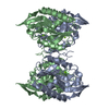
- Assembly
| Deposited unit | 
| ||||||||
|---|---|---|---|---|---|---|---|---|---|
| 1 | 
| ||||||||
| Unit cell |
|
-Components
| #1: Protein | Mass: 20657.887 Da / Num. of mol.: 2 Source method: isolated from a genetically manipulated source Source: (gene. exp.) References: UniProt: P0A9M2, hypoxanthine phosphoribosyltransferase #2: Chemical | ChemComp-5GP / |   Mass: 363.221 Da / Num. of mol.: 1 / Source method: obtained synthetically / Formula: C10H14N5O8P Mass: 363.221 Da / Num. of mol.: 1 / Source method: obtained synthetically / Formula: C10H14N5O8P#3: Chemical | ChemComp-N / |   Type: RNA linking / Mass: 214.110 Da / Num. of mol.: 1 / Source method: obtained synthetically / Formula: C5H11O7P Type: RNA linking / Mass: 214.110 Da / Num. of mol.: 1 / Source method: obtained synthetically / Formula: C5H11O7P#4: Water | ChemComp-HOH / |  Mass: 18.015 Da / Num. of mol.: 114 / Source method: isolated from a natural source / Formula: H2O Mass: 18.015 Da / Num. of mol.: 114 / Source method: isolated from a natural source / Formula: H2O |
|---|
-Experimental details
-Experiment
| Experiment | Method: X-RAY DIFFRACTION / Number of used crystals: 1 |
|---|
- Sample preparation
| Crystal | Density Matthews: 4.14 Å3/Da / Density % sol: 70.26 % | |||||||||||||||||||||||||||||||||||||||||||||||||
|---|---|---|---|---|---|---|---|---|---|---|---|---|---|---|---|---|---|---|---|---|---|---|---|---|---|---|---|---|---|---|---|---|---|---|---|---|---|---|---|---|---|---|---|---|---|---|---|---|---|---|
| Crystal grow | Temperature: 293 K / Method: vapor diffusion, hanging drop / pH: 7.5 Details: Hepes, sodium citrate, pH 7.5, VAPOR DIFFUSION, HANGING DROP, temperature 293K | |||||||||||||||||||||||||||||||||||||||||||||||||
| Crystal grow | *PLUS Temperature: 20 ℃ / pH: 7.4 | |||||||||||||||||||||||||||||||||||||||||||||||||
| Components of the solutions | *PLUS
|
-Data collection
| Diffraction | Mean temperature: 100 K |
|---|---|
| Diffraction source | Source: SYNCHROTRON / Site: APS  / Beamline: 14-BM-D / Wavelength: 1 Å / Beamline: 14-BM-D / Wavelength: 1 Å |
| Detector | Type: ADSC QUANTUM 1 / Detector: CCD / Date: Dec 20, 1999 / Details: mirrors |
| Radiation | Monochromator: mirror / Protocol: SINGLE WAVELENGTH / Monochromatic (M) / Laue (L): M / Scattering type: x-ray |
| Radiation wavelength | Wavelength: 1 Å / Relative weight: 1 |
| Reflection | Resolution: 2.8→50 Å / Num. all: 15697 / Num. obs: 45289 / % possible obs: 88.9 % / Observed criterion σ(F): 0 / Observed criterion σ(I): 0 / Redundancy: 2.9 % / Biso Wilson estimate: 38 Å2 / Rmerge(I) obs: 0.072 / Net I/σ(I): 9.1 |
| Reflection shell | Resolution: 2.8→2.9 Å / Redundancy: 1.2 % / Rmerge(I) obs: 0.294 / Mean I/σ(I) obs: 4.1 / Num. unique all: 1388 / % possible all: 89.8 |
| Reflection | *PLUS Lowest resolution: 50 Å / Num. obs: 15697 / Num. measured all: 45289 / Rmerge(I) obs: 0.072 |
| Reflection shell | *PLUS Highest resolution: 2.8 Å / Lowest resolution: 2.9 Å / % possible obs: 89.8 % / Rmerge(I) obs: 0.294 |
- Processing
| Software |
| ||||||||||||||||||||
|---|---|---|---|---|---|---|---|---|---|---|---|---|---|---|---|---|---|---|---|---|---|
| Refinement | Method to determine structure: MOLECULAR REPLACEMENT Starting model: T.foetus HPRT Resolution: 2.8→50 Å / Cross valid method: USED THROUGHOUT / σ(F): 0 / σ(I): 0 / Stereochemistry target values: Engh & Huber / Details: simulated annealing refinement
| ||||||||||||||||||||
| Displacement parameters | Biso mean: 38.7 Å2
| ||||||||||||||||||||
| Refine analyze |
| ||||||||||||||||||||
| Refinement step | Cycle: LAST / Resolution: 2.8→50 Å
| ||||||||||||||||||||
| Refine LS restraints |
| ||||||||||||||||||||
| LS refinement shell | Resolution: 2.8→2.9 Å / Rfactor Rfree error: 0.012
| ||||||||||||||||||||
| Refinement | *PLUS % reflection Rfree: 10 % / Rfactor all: 0.199 / Rfactor Rfree: 0.244 / Rfactor Rwork: 0.199 | ||||||||||||||||||||
| Solvent computation | *PLUS | ||||||||||||||||||||
| Displacement parameters | *PLUS | ||||||||||||||||||||
| Refine LS restraints | *PLUS Type: c_angle_deg / Dev ideal: 1.38 | ||||||||||||||||||||
| LS refinement shell | *PLUS Rfactor Rfree: 0.3338 / Rfactor Rwork: 0.2748 |
