 Movie
Movie Controller

[English] 日本語
 Yorodumi
- PDB-1g7k: CRYSTAL STRUCTURE OF DSRED, A RED FLUORESCENT PROTEIN FROM DISCOS... -
+ Open data

- Basic information
| Entry | Database: PDB / ID: 1g7k | |||||||||
|---|---|---|---|---|---|---|---|---|---|---|
| Title | CRYSTAL STRUCTURE OF DSRED, A RED FLUORESCENT PROTEIN FROM DISCOSOMA SP. RED | |||||||||
 Components Components | FLUORESCENT PROTEIN FP583 | |||||||||
 Keywords Keywords | LUMINESCENT PROTEIN / Fluorescent Protein / GFP / Coral / Red / Beta Barrel / Chromophore / dsred / drFP583 | |||||||||
| Function / homology |  Function and homology information Function and homology information | |||||||||
| Biological species |  Discosoma sp. (sea anemone) Discosoma sp. (sea anemone) | |||||||||
| Method |  X-RAY DIFFRACTION / SYNCHROTRON / MAD / Resolution: 2 Å X-RAY DIFFRACTION / SYNCHROTRON / MAD / Resolution: 2 Å | |||||||||
 Authors Authors | Yarbrough, D. / Wachter, R.M. / Kallio, K. / Matz, M.V. / Remington, S.J. | |||||||||
 Citation Citation | Journal: Proc.Natl.Acad.Sci.USA / Year: 2001 Title: Refined crystal structure of DsRed, a red fluorescent protein from coral, at 2.0-A resolution. Authors: Yarbrough, D. / Wachter, R.M. / Kallio, K. / Matz, M.V. / Remington, S.J. #1: Journal: Nat.Biotechnol. / Year: 1999Title: Fluorescent Proteins from Nonbioluminescent Anthozoa Species Authors: Matz, M.V. / Fradkov, A.F. / Labas, Y.A. / Savitsky, A.P. / Zaraisky, A.G. / Markelov, M.L. / Lukyanov, S.A. #2: Journal: Proc.Natl.Acad.Sci.USA / Year: 2000Title: The Structure of the Chromophore within DsRed, a Red Fluorescent Protein from Coral Authors: Gross, L.A. / Baird, G.S. / Hoffman, R.C. / Baldridge, K.K. / Tsien, R.Y. | |||||||||
| History |
|
- Structure visualization
| Structure viewer | Molecule: MolmilJmol/JSmol |
|---|
- Downloads & links
-Download
| PDBx/mmCIF format | 1g7k.cif.gz | 196.9 KB | Display | PDBx/mmCIF format |
|---|---|---|---|---|
| PDB format | pdb1g7k.ent.gz | 158.8 KB | Display | PDB format |
| PDBx/mmJSON format | 1g7k.json.gz | Tree view | PDBx/mmJSON format | |
| Others |  Other downloads Other downloads |
-Validation report
| Summary document | 1g7k_validation.pdf.gz | 455.2 KB | Display | wwPDB validaton report |
|---|---|---|---|---|
| Full document | 1g7k_full_validation.pdf.gz | 495.7 KB | Display | |
| Data in XML | 1g7k_validation.xml.gz | 42.1 KB | Display | |
| Data in CIF | 1g7k_validation.cif.gz | 58 KB | Display | |
| Arichive directory | https://data.pdbj.org/pub/pdb/validation_reports/g7/1g7kftp://data.pdbj.org/pub/pdb/validation_reports/g7/1g7k | HTTPS FTP |
-Related structure data
| Similar structure data |
|---|


-Links
 PDBj
PDBj

- Assembly
| Deposited unit | 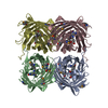
| ||||||||
|---|---|---|---|---|---|---|---|---|---|
| 1 |
| ||||||||
| Unit cell |
| ||||||||
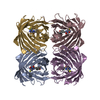
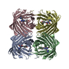
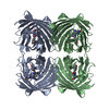
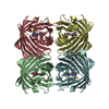
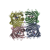
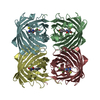
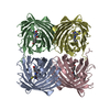
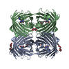
| Details | the full biological assembly is the homotetramer in the asymmetric unit |
-Components
| #1: Protein | Mass: 27551.205 Da / Num. of mol.: 4 / Mutation: RESIDUES 66-68 REPLACED BY CRO Source method: isolated from a genetically manipulated source Source: (gene. exp.) Discosoma sp. (sea anemone) / Gene: DRFP583 / Plasmid: PQE-30 / Production host:  #2: Water | ChemComp-HOH / |  Mass: 18.015 Da / Num. of mol.: 418 / Source method: isolated from a natural source / Formula: H2O Mass: 18.015 Da / Num. of mol.: 418 / Source method: isolated from a natural source / Formula: H2OHas protein modification | Y | Sequence details | RESIDUES GLN 66, TYR 67, GLY 68 ARE MODIFIED TO MAKE THE CHROMOPHOR | |
|---|
-Experimental details
-Experiment
| Experiment | Method: X-RAY DIFFRACTION / Number of used crystals: 1 |
|---|
- Sample preparation
| Crystal | Density Matthews: 2.02 Å3/Da / Density % sol: 39.24 % | ||||||||||||||||||||||||||||||||||||||||||
|---|---|---|---|---|---|---|---|---|---|---|---|---|---|---|---|---|---|---|---|---|---|---|---|---|---|---|---|---|---|---|---|---|---|---|---|---|---|---|---|---|---|---|---|
| Crystal grow | Temperature: 298 K / Method: vapor diffusion, hanging drop / pH: 7.8 Details: PEG 4000, Tris, Beta-Mercaptoethanol, pH 7.8, VAPOR DIFFUSION, HANGING DROP, temperature 298K | ||||||||||||||||||||||||||||||||||||||||||
| Crystal grow | *PLUS pH: 7.9 / Details: used microseeding | ||||||||||||||||||||||||||||||||||||||||||
| Components of the solutions | *PLUS
|
-Data collection
| Diffraction | Mean temperature: 100 K | ||||||||||||
|---|---|---|---|---|---|---|---|---|---|---|---|---|---|
| Diffraction source | Source: SYNCHROTRON / Site: SSRL  / Beamline: BL9-2 / Wavelength: 0.97887,0.97868,0.95373 / Beamline: BL9-2 / Wavelength: 0.97887,0.97868,0.95373 | ||||||||||||
| Detector | Type: ADSC QUANTUM 4 / Detector: CCD / Date: Apr 21, 1999 / Details: mirrors | ||||||||||||
| Radiation | Monochromator: Double-Crystal Si 111 crystals / Protocol: MAD / Monochromatic (M) / Laue (L): M / Scattering type: x-ray | ||||||||||||
| Radiation wavelength |
| ||||||||||||
| Reflection | Resolution: 2→24.1 Å / Num. all: 56056 / Num. obs: 56056 / % possible obs: 94.8 % / Observed criterion σ(F): 2 / Observed criterion σ(I): 2 / Redundancy: 4.1 % / Biso Wilson estimate: 10.2 Å2 / Rmerge(I) obs: 0.026 / Net I/σ(I): 18.6 | ||||||||||||
| Reflection shell | Resolution: 2→2.1 Å / Redundancy: 4.1 % / Rmerge(I) obs: 0.043 / % possible all: 95.1 | ||||||||||||
| Reflection | *PLUS Highest resolution: 2 Å / Lowest resolution: 25.1 Å / Num. obs: 56118 / Num. measured all: 232348 / Rmerge(I) obs: 0.027 | ||||||||||||
| Reflection shell | *PLUS Lowest resolution: 2.11 Å / Rmerge(I) obs: 0.044 |
- Processing
| Software |
| |||||||||||||||||||||||||
|---|---|---|---|---|---|---|---|---|---|---|---|---|---|---|---|---|---|---|---|---|---|---|---|---|---|---|
| Refinement | Method to determine structure: MAD / Resolution: 2→30 Å / σ(F): 0 / σ(I): 0 / Stereochemistry target values: TNT
| |||||||||||||||||||||||||
| Refinement step | Cycle: LAST / Resolution: 2→30 Å
| |||||||||||||||||||||||||
| Refine LS restraints |
| |||||||||||||||||||||||||
| Software | *PLUS Name: TNT / Classification: refinement | |||||||||||||||||||||||||
| Refinement | *PLUS Highest resolution: 2 Å / Lowest resolution: 25.1 Å / σ(F): 0 / Rfactor obs: 0.162 | |||||||||||||||||||||||||
| Solvent computation | *PLUS | |||||||||||||||||||||||||
| Displacement parameters | *PLUS |
