 Movie
Movie Controller


+ Open data

- Basic information
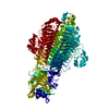
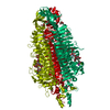
| Entry | Database: PDB / ID: 1g5g | |||||||||
|---|---|---|---|---|---|---|---|---|---|---|
| Title | FRAGMENT OF FUSION PROTEIN FROM NEWCASTLE DISEASE VIRUS | |||||||||
 Components Components | Fusion glycoprotein F0 | |||||||||
 Keywords Keywords | VIRAL PROTEIN / fusion protein / NDV / Newcastle disease virus / paramyxovirus | |||||||||
| Function / homology |  Function and homology information Function and homology informationfusion of virus membrane with host plasma membrane / viral envelope / symbiont entry into host cell / host cell plasma membrane / virion membrane Similarity search - Function | |||||||||
| Biological species |  Avian orthoavulavirus 1 Avian orthoavulavirus 1 | |||||||||
| Method |  X-RAY DIFFRACTION / SYNCHROTRON / SIR, phase extension / Resolution: 3.3 Å X-RAY DIFFRACTION / SYNCHROTRON / SIR, phase extension / Resolution: 3.3 Å | |||||||||
 Authors Authors | Lawrence, M.C. / Smith, B.J. | |||||||||
 Citation Citation | Journal: Structure / Year: 2001 Title: The structure of the fusion glycoprotein of Newcastle disease virus suggests a novel paradigm for the molecular mechanism of membrane fusion. Authors: Chen, L. / Gorman, J.J. / McKimm-Breschkin, J. / Lawrence, L.J. / Tulloch, P.A. / Smith, B.J. / Colman, P.M. / Lawrence, M.C. | |||||||||
| History |
|
- Structure visualization
| Structure viewer | Molecule: MolmilJmol/JSmol |
|---|
- Downloads & links
-Download
| PDBx/mmCIF format | 1g5g.cif.gz | 413.5 KB | Display | PDBx/mmCIF format |
|---|---|---|---|---|
| PDB format | pdb1g5g.ent.gz | 336.8 KB | Display | PDB format |
| PDBx/mmJSON format | 1g5g.json.gz | Tree view | PDBx/mmJSON format | |
| Others |  Other downloads Other downloads |
-Validation report
| Arichive directory | https://data.pdbj.org/pub/pdb/validation_reports/g5/1g5gftp://data.pdbj.org/pub/pdb/validation_reports/g5/1g5g | HTTPS FTP |
|---|
-Related structure data
| Similar structure data |
|---|
-Links
 PDBj
PDBj

- Assembly
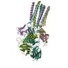
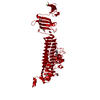
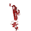
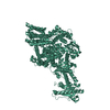
| Deposited unit | 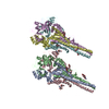
| ||||||||
|---|---|---|---|---|---|---|---|---|---|
| 1 | 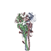
| ||||||||
| 2 | 
| ||||||||
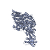
| Unit cell |
|
-Components
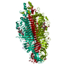
| #1: Protein | Mass: 51086.871 Da / Num. of mol.: 6 Fragment: FULL-LENGTH ECTODOMAIN (RESIDUES L32-R501) FOLLOWED BY C-MYC PURIFICATION TAG Source method: isolated from a genetically manipulated source Source: (gene. exp.) Avian orthoavulavirus 1 / Genus: AvulavirusStrain: Newcastle disease virus strain Queensland/66, V4 ISOLATE References: UniProt: A9LSB1, UniProt: P35936*PLUS #2: Polysaccharide | 2-acetamido-2-deoxy-beta-D-glucopyranose-(1-4)-2-acetamido-2-deoxy-beta-D-glucopyranose Source method: isolated from a genetically manipulated source #3: Polysaccharide | beta-D-mannopyranose-(1-4)-2-acetamido-2-deoxy-beta-D-glucopyranose-(1-4)-2-acetamido-2-deoxy-beta- ...beta-D-mannopyranose-(1-4)-2-acetamido-2-deoxy-beta-D-glucopyranose-(1-4)-2-acetamido-2-deoxy-beta-D-glucopyranose | Source method: isolated from a genetically manipulated source #4: Sugar | ChemComp-NAG /   Type: D-saccharide, beta linking / Mass: 221.208 Da / Num. of mol.: 4 Type: D-saccharide, beta linking / Mass: 221.208 Da / Num. of mol.: 4Source method: isolated from a genetically manipulated source Formula: C8H15NO6 Has protein modification | Y | |
|---|
-Experimental details
-Experiment
| Experiment | Method: X-RAY DIFFRACTION / Number of used crystals: 15 |
|---|
- Sample preparation
| Crystal |
| |||||||||||||||
|---|---|---|---|---|---|---|---|---|---|---|---|---|---|---|---|---|
| Crystal grow |
| |||||||||||||||
| Crystal grow | *PLUS Method: unknown / Details: unpublished data |
-Data collection
| Diffraction |
| |||||||||||||||||||||||||||||||||||
|---|---|---|---|---|---|---|---|---|---|---|---|---|---|---|---|---|---|---|---|---|---|---|---|---|---|---|---|---|---|---|---|---|---|---|---|---|
| Diffraction source |
| |||||||||||||||||||||||||||||||||||
| Detector |
| |||||||||||||||||||||||||||||||||||
| Radiation | Protocol: SINGLE WAVELENGTH / Monochromatic (M) / Laue (L): M / Scattering type: x-ray | |||||||||||||||||||||||||||||||||||
| Radiation wavelength |
| |||||||||||||||||||||||||||||||||||
| Reflection | Resolution: 3.3→25 Å / Num. all: 76017 / Num. obs: 73130 / % possible obs: 96.2 % / Observed criterion σ(F): 0 / Observed criterion σ(I): 0 / Redundancy: 6.1 % / Rmerge(I) obs: 0.22 / Net I/σ(I): 7.04 | |||||||||||||||||||||||||||||||||||
| Reflection shell | Resolution: 3.3→3.42 Å / Redundancy: 2.3 % / Rmerge(I) obs: 0.812 / Num. unique all: 7502 / % possible all: 87.2 | |||||||||||||||||||||||||||||||||||
| Reflection | *PLUS Num. measured all: 438164 | |||||||||||||||||||||||||||||||||||
| Reflection shell | *PLUS % possible obs: 87.2 % / Rmerge(I) obs: 0.88 / Mean I/σ(I) obs: 1.2 |

- Processing
| Software |
| ||||||||||||||||||||
|---|---|---|---|---|---|---|---|---|---|---|---|---|---|---|---|---|---|---|---|---|---|
| Refinement | Method to determine structure: SIR, phase extension / Resolution: 3.3→8 Å / σ(F): 0 / σ(I): 0 / Stereochemistry target values: Engh & Huber
| ||||||||||||||||||||
| Refinement step | Cycle: LAST / Resolution: 3.3→8 Å
| ||||||||||||||||||||
| Refine LS restraints |
| ||||||||||||||||||||
| Software | *PLUS Name: X-PLOR / Version: 3.851 / Classification: refinement | ||||||||||||||||||||
| Refinement | *PLUS σ(F): 0 / Rfactor obs: 0.224 | ||||||||||||||||||||
| Solvent computation | *PLUS | ||||||||||||||||||||
| Displacement parameters | *PLUS | ||||||||||||||||||||
| Refine LS restraints | *PLUS Type: x_angle_deg / Dev ideal: 1.5 |
