 Movie
Movie Controller

[English] 日本語
 Yorodumi
- PDB-1g3m: CRYSTAL STRUCTURE OF HUMAN ESTROGEN SULFOTRANSFERASE IN COMPLEX W... -
+ Open data

- Basic information
| Entry | Database: PDB / ID: 1g3m | ||||||
|---|---|---|---|---|---|---|---|
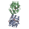
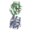
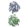
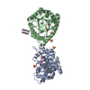
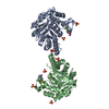
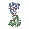
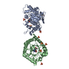
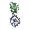
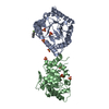
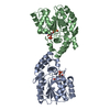
| Title | CRYSTAL STRUCTURE OF HUMAN ESTROGEN SULFOTRANSFERASE IN COMPLEX WITH IN-ACTIVE COFACTOR PAP AND 3,5,3',5'-TETRACHLORO-BIPHENYL-4,4'-DIOL | ||||||
 Components Components | ESTROGEN SULFOTRANSFERASE | ||||||
 Keywords Keywords | TRANSFERASE / estrogen / sulfotransferase / PCB / human | ||||||
| Function / homology |  Function and homology information Function and homology informationestrone sulfotransferase / estrone sulfotransferase activity / estrogen catabolic process / flavonol 3-sulfotransferase activity / steroid sulfotransferase activity / aryl sulfotransferase activity / Cytosolic sulfonation of small molecules / 3'-phosphoadenosine 5'-phosphosulfate metabolic process / sulfation / sulfotransferase activity ...estrone sulfotransferase / estrone sulfotransferase activity / estrogen catabolic process / flavonol 3-sulfotransferase activity / steroid sulfotransferase activity / aryl sulfotransferase activity / Cytosolic sulfonation of small molecules / 3'-phosphoadenosine 5'-phosphosulfate metabolic process / sulfation / sulfotransferase activity / ethanol catabolic process / Paracetamol ADME / estrogen metabolic process / steroid metabolic process / positive regulation of fat cell differentiation / steroid binding / nuclear membrane / cytosol / cytoplasm Similarity search - Function | ||||||
| Biological species |  Homo sapiens (human) Homo sapiens (human) | ||||||
| Method |  X-RAY DIFFRACTION / MOLECULAR REPLACEMENT / Resolution: 1.7 Å X-RAY DIFFRACTION / MOLECULAR REPLACEMENT / Resolution: 1.7 Å | ||||||
 Authors Authors | Shevtsov, S. / Petrochenko, E.V. / Negishi, M. / Pedersen, L.C. | ||||||
 Citation Citation | Journal: Environ.Health Perspect. / Year: 2003 Title: Crystallographic analysis of a hydroxylated polychlorinated biphenyl (OH-PCB) bound to the catalytic estrogen binding site of human estrogen sulfotransferase. Authors: Shevtsov, S. / Petrotchenko, E.V. / Pedersen, L.C. / Negishi, M. | ||||||
| History |
|
- Structure visualization
| Structure viewer | Molecule: MolmilJmol/JSmol |
|---|
- Downloads & links
-Download
| PDBx/mmCIF format | 1g3m.cif.gz | 147.4 KB | Display | PDBx/mmCIF format |
|---|---|---|---|---|
| PDB format | pdb1g3m.ent.gz | 113.8 KB | Display | PDB format |
| PDBx/mmJSON format | 1g3m.json.gz | Tree view | PDBx/mmJSON format | |
| Others |  Other downloads Other downloads |
-Validation report
| Arichive directory | https://data.pdbj.org/pub/pdb/validation_reports/g3/1g3mftp://data.pdbj.org/pub/pdb/validation_reports/g3/1g3m | HTTPS FTP |
|---|
-Related structure data
| Similar structure data |
|---|
-Links
 PDBj
PDBj
- Assembly
| Deposited unit | 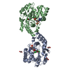
| ||||||||
|---|---|---|---|---|---|---|---|---|---|
| 1 |
| ||||||||
| Unit cell |
| ||||||||
| Details | The asymmetric unit represents the proposed biological dimer. |
-Components
| #1: Protein | Mass: 35176.375 Da / Num. of mol.: 2 Source method: isolated from a genetically manipulated source Source: (gene. exp.) Homo sapiens (human) / Gene: STE / Plasmid: PGEX4T3 / Species (production host): Escherichia coli / Production host:  #2: Chemical |   Type: RNA linking / Mass: 427.201 Da / Num. of mol.: 2 / Source method: obtained synthetically / Formula: C10H15N5O10P2 Type: RNA linking / Mass: 427.201 Da / Num. of mol.: 2 / Source method: obtained synthetically / Formula: C10H15N5O10P2#3: Chemical |   Mass: 323.987 Da / Num. of mol.: 2 / Source method: obtained synthetically / Formula: C12H6Cl4O2 Mass: 323.987 Da / Num. of mol.: 2 / Source method: obtained synthetically / Formula: C12H6Cl4O2#4: Chemical | ChemComp-EDO /   Mass: 62.068 Da / Num. of mol.: 4 / Source method: obtained synthetically / Formula: C2H6O2 Mass: 62.068 Da / Num. of mol.: 4 / Source method: obtained synthetically / Formula: C2H6O2#5: Water | ChemComp-HOH / |  Mass: 18.015 Da / Num. of mol.: 603 / Source method: isolated from a natural source / Formula: H2O Mass: 18.015 Da / Num. of mol.: 603 / Source method: isolated from a natural source / Formula: H2O |
|---|
-Experimental details
-Experiment
| Experiment | Method: X-RAY DIFFRACTION / Number of used crystals: 1 |
|---|
- Sample preparation
| Crystal | Density Matthews: 2.66 Å3/Da / Density % sol: 53.77 % | |||||||||||||||||||||||||||||||||||||||||||||||||
|---|---|---|---|---|---|---|---|---|---|---|---|---|---|---|---|---|---|---|---|---|---|---|---|---|---|---|---|---|---|---|---|---|---|---|---|---|---|---|---|---|---|---|---|---|---|---|---|---|---|---|
| Crystal grow | Temperature: 295 K / Method: vapor diffusion, hanging drop / pH: 6 Details: 0.1M MES 22% PEG8000 , pH 6.0, VAPOR DIFFUSION, HANGING DROP, temperature 295K | |||||||||||||||||||||||||||||||||||||||||||||||||
| Crystal grow | *PLUS Temperature: 20 ℃ / pH: 7.5 / Method: vapor diffusion, sitting drop | |||||||||||||||||||||||||||||||||||||||||||||||||
| Components of the solutions | *PLUS
|
-Data collection
| Diffraction | Mean temperature: 93 K |
|---|---|
| Diffraction source | Source: ROTATING ANODE / Type: RIGAKU RU300 / Wavelength: 1.542 Å |
| Detector | Type: RIGAKU RAXIS IV / Detector: IMAGE PLATE / Date: Sep 15, 2000 / Details: mirrors |
| Radiation | Protocol: SINGLE WAVELENGTH / Monochromatic (M) / Laue (L): M / Scattering type: x-ray |
| Radiation wavelength | Wavelength: 1.542 Å / Relative weight: 1 |
| Reflection | Resolution: 1.7→25 Å / Num. obs: 81460 / % possible obs: 92.6 % / Observed criterion σ(I): -3 / Redundancy: 2 % / Biso Wilson estimate: 18.4 Å2 / Rsym value: 0.058 / Net I/σ(I): 8.39 |
| Reflection shell | Resolution: 1.7→1.76 Å / Redundancy: 1.37 % / Mean I/σ(I) obs: 1.94 / Rsym value: 0.262 / % possible all: 82.9 |
| Reflection | *PLUS Lowest resolution: 50 Å / Num. obs: 75359 / Num. measured all: 151066 / Rmerge(I) obs: 0.058 |
| Reflection shell | *PLUS % possible obs: 82.9 % / Rmerge(I) obs: 0.262 / Mean I/σ(I) obs: 1.9 |
- Processing
| Software |
| ||||||||||||||||||||||||||||||||||||||||||||||||||||||||||||||||||||||||||||||||
|---|---|---|---|---|---|---|---|---|---|---|---|---|---|---|---|---|---|---|---|---|---|---|---|---|---|---|---|---|---|---|---|---|---|---|---|---|---|---|---|---|---|---|---|---|---|---|---|---|---|---|---|---|---|---|---|---|---|---|---|---|---|---|---|---|---|---|---|---|---|---|---|---|---|---|---|---|---|---|---|---|---|
| Refinement | Method to determine structure: MOLECULAR REPLACEMENT Starting model: HUMAN EST Resolution: 1.7→24.53 Å / Rfactor Rfree error: 0.004 / Data cutoff high absF: 644110.07 / Data cutoff low absF: 0 / Isotropic thermal model: RESTRAINED / Cross valid method: THROUGHOUT / σ(F): 0
| ||||||||||||||||||||||||||||||||||||||||||||||||||||||||||||||||||||||||||||||||
| Solvent computation | Solvent model: FLAT MODEL / Bsol: 51.22 Å2 / ksol: 0.369 e/Å3 | ||||||||||||||||||||||||||||||||||||||||||||||||||||||||||||||||||||||||||||||||
| Displacement parameters | Biso mean: 21.4 Å2
| ||||||||||||||||||||||||||||||||||||||||||||||||||||||||||||||||||||||||||||||||
| Refine analyze |
| ||||||||||||||||||||||||||||||||||||||||||||||||||||||||||||||||||||||||||||||||
| Refinement step | Cycle: LAST / Resolution: 1.7→24.53 Å
| ||||||||||||||||||||||||||||||||||||||||||||||||||||||||||||||||||||||||||||||||
| Refine LS restraints |
| ||||||||||||||||||||||||||||||||||||||||||||||||||||||||||||||||||||||||||||||||
| LS refinement shell | Resolution: 1.7→1.81 Å / Rfactor Rfree error: 0.012 / Total num. of bins used: 6
| ||||||||||||||||||||||||||||||||||||||||||||||||||||||||||||||||||||||||||||||||
| Xplor file |
| ||||||||||||||||||||||||||||||||||||||||||||||||||||||||||||||||||||||||||||||||
| Refinement | *PLUS Highest resolution: 1.7 Å / Lowest resolution: 50 Å / % reflection Rfree: 5 % | ||||||||||||||||||||||||||||||||||||||||||||||||||||||||||||||||||||||||||||||||
| Solvent computation | *PLUS | ||||||||||||||||||||||||||||||||||||||||||||||||||||||||||||||||||||||||||||||||
| Displacement parameters | *PLUS | ||||||||||||||||||||||||||||||||||||||||||||||||||||||||||||||||||||||||||||||||
| Refine LS restraints | *PLUS
|
