 Movie
Movie Controller

[English] 日本語
 Yorodumi
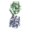
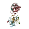
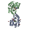
- PDB-1hy3: CRYSTAL STRUCTURE OF HUMAN ESTROGEN SULFOTRANSFERASE V269E MUTANT... -
+ Open data

- Basic information
| Entry | Database: PDB / ID: 1hy3 | ||||||
|---|---|---|---|---|---|---|---|
| Title | CRYSTAL STRUCTURE OF HUMAN ESTROGEN SULFOTRANSFERASE V269E MUTANT IN THE PRESENCE OF PAPS | ||||||
 Components Components | ESTROGEN SULFOTRANSFERASE | ||||||
 Keywords Keywords | TRANSFERASE / ESTROGEN / SULFOTRANSFERASE / PAPS / HUMAN | ||||||
| Function / homology |  Function and homology information Function and homology informationestrone sulfotransferase / estrone sulfotransferase activity / estrogen catabolic process / flavonol 3-sulfotransferase activity / steroid sulfotransferase activity / aryl sulfotransferase activity / Cytosolic sulfonation of small molecules / 3'-phosphoadenosine 5'-phosphosulfate metabolic process / sulfation / sulfotransferase activity ...estrone sulfotransferase / estrone sulfotransferase activity / estrogen catabolic process / flavonol 3-sulfotransferase activity / steroid sulfotransferase activity / aryl sulfotransferase activity / Cytosolic sulfonation of small molecules / 3'-phosphoadenosine 5'-phosphosulfate metabolic process / sulfation / sulfotransferase activity / ethanol catabolic process / Paracetamol ADME / estrogen metabolic process / steroid metabolic process / positive regulation of fat cell differentiation / steroid binding / nuclear membrane / cytoplasm / cytosol Similarity search - Function | ||||||
| Biological species |  Homo sapiens (human) Homo sapiens (human) | ||||||
| Method |  X-RAY DIFFRACTION / MOLECULAR REPLACEMENT / Resolution: 1.8 Å X-RAY DIFFRACTION / MOLECULAR REPLACEMENT / Resolution: 1.8 Å | ||||||
 Authors Authors | Pedersen, L.C. / Petrochenko, E.V. / Shevtsov, S. / Negishi, M. | ||||||
 Citation Citation | Journal: J.Biol.Chem. / Year: 2002 Title: Crystal structure of the human estrogen sulfotransferase-PAPS complex: evidence for catalytic role of Ser137 in the sulfuryl transfer reaction. Authors: Pedersen, L.C. / Petrotchenko, E. / Shevtsov, S. / Negishi, M. | ||||||
| History |
|
- Structure visualization
| Structure viewer | Molecule: MolmilJmol/JSmol |
|---|
- Downloads & links
-Download
| PDBx/mmCIF format | 1hy3.cif.gz | 141.9 KB | Display | PDBx/mmCIF format |
|---|---|---|---|---|
| PDB format | pdb1hy3.ent.gz | 110.4 KB | Display | PDB format |
| PDBx/mmJSON format | 1hy3.json.gz | Tree view | PDBx/mmJSON format | |
| Others |  Other downloads Other downloads |
-Validation report
| Arichive directory | https://data.pdbj.org/pub/pdb/validation_reports/hy/1hy3ftp://data.pdbj.org/pub/pdb/validation_reports/hy/1hy3 | HTTPS FTP |
|---|
-Related structure data
| Related structure data | |
|---|---|
| Similar structure data |


-Links
 PDBj
PDBj
- Assembly
| Deposited unit | 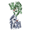
| ||||||||
|---|---|---|---|---|---|---|---|---|---|
| 1 |
| ||||||||
| Unit cell |
| ||||||||
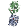
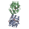
| Details | ASYMMETRIC UNIT MOST LIKELY REPRESENTS FUNCTIONAL DIMER |
-Components
| #1: Protein | Mass: 35206.359 Da / Num. of mol.: 2 / Mutation: V269E Source method: isolated from a genetically manipulated source Source: (gene. exp.) Homo sapiens (human) / Gene: STE / Plasmid: PGEX4T3 / Species (production host): Escherichia coli / Production host:  #2: Chemical | 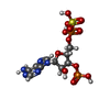  Mass: 507.264 Da / Num. of mol.: 2 / Source method: obtained synthetically / Formula: C10H15N5O13P2S Mass: 507.264 Da / Num. of mol.: 2 / Source method: obtained synthetically / Formula: C10H15N5O13P2S#3: Water | ChemComp-HOH / |  Mass: 18.015 Da / Num. of mol.: 450 / Source method: isolated from a natural source / Formula: H2O Mass: 18.015 Da / Num. of mol.: 450 / Source method: isolated from a natural source / Formula: H2O |
|---|
-Experimental details
-Experiment
| Experiment | Method: X-RAY DIFFRACTION / Number of used crystals: 1 |
|---|
- Sample preparation
| Crystal | Density Matthews: 2.68 Å3/Da / Density % sol: 54.11 % | |||||||||||||||||||||||||||||||||||||||||||||||||
|---|---|---|---|---|---|---|---|---|---|---|---|---|---|---|---|---|---|---|---|---|---|---|---|---|---|---|---|---|---|---|---|---|---|---|---|---|---|---|---|---|---|---|---|---|---|---|---|---|---|---|
| Crystal grow | Temperature: 298 K / Method: vapor diffusion, hanging drop / pH: 6 Details: MES, PEG 8K, PAP, pH 6.0, VAPOR DIFFUSION, HANGING DROP, temperature 298K | |||||||||||||||||||||||||||||||||||||||||||||||||
| Crystal grow | *PLUS Temperature: 20 ℃ / pH: 7.2 / Method: vapor diffusion, sitting drop | |||||||||||||||||||||||||||||||||||||||||||||||||
| Components of the solutions | *PLUS
|
-Data collection
| Diffraction | Mean temperature: 93 K |
|---|---|
| Diffraction source | Source: ROTATING ANODE / Type: RIGAKU RU300 / Wavelength: 1.5418 Å |
| Detector | Type: RIGAKU RAXIS IV / Detector: IMAGE PLATE / Date: Aug 23, 2000 / Details: MIRRORS |
| Radiation | Protocol: SINGLE WAVELENGTH / Monochromatic (M) / Laue (L): M / Scattering type: x-ray |
| Radiation wavelength | Wavelength: 1.5418 Å / Relative weight: 1 |
| Reflection | Resolution: 1.8→25 Å / Num. all: 63064 / Num. obs: 62882 / % possible obs: 91.8 % / Observed criterion σ(F): 0 / Observed criterion σ(I): -3 / Redundancy: 2.9 % / Biso Wilson estimate: 21 Å2 / Rmerge(I) obs: 0.049 / Rsym value: 0.049 / Net I/σ(I): 19.6 |
| Reflection shell | Resolution: 1.8→1.86 Å / Redundancy: 1.7 % / Rmerge(I) obs: 0.229 / Mean I/σ(I) obs: 2.22 / Num. unique all: 3569 / % possible all: 52.1 |
| Reflection | *PLUS Num. obs: 63064 / Num. measured all: 182966 / Rmerge(I) obs: 0.049 |
| Reflection shell | *PLUS % possible obs: 52.1 % / Rmerge(I) obs: 0.229 / Mean I/σ(I) obs: 2.2 |
- Processing
| Software |
| ||||||||||||||||||||||||||||||||||||
|---|---|---|---|---|---|---|---|---|---|---|---|---|---|---|---|---|---|---|---|---|---|---|---|---|---|---|---|---|---|---|---|---|---|---|---|---|---|
| Refinement | Method to determine structure: MOLECULAR REPLACEMENT Starting model: HUMAN EST Resolution: 1.8→22.54 Å / Rfactor Rfree error: 0.004 / Data cutoff high absF: 494998.12 / Data cutoff low absF: 0 / Isotropic thermal model: RESTRAINED / Cross valid method: THROUGHOUT / σ(F): 0 / σ(I): 0 / Stereochemistry target values: CNS target values
| ||||||||||||||||||||||||||||||||||||
| Solvent computation | Solvent model: FLAT MODEL / Bsol: 39.4 Å2 / ksol: 0.346 e/Å3 | ||||||||||||||||||||||||||||||||||||
| Displacement parameters | Biso mean: 27.3 Å2 | ||||||||||||||||||||||||||||||||||||
| Refine analyze |
| ||||||||||||||||||||||||||||||||||||
| Refinement step | Cycle: LAST / Resolution: 1.8→22.54 Å
| ||||||||||||||||||||||||||||||||||||
| Refine LS restraints |
| ||||||||||||||||||||||||||||||||||||
| LS refinement shell | Resolution: 1.8→1.91 Å / Rfactor Rfree error: 0.016 / Total num. of bins used: 6
| ||||||||||||||||||||||||||||||||||||
| Xplor file |
| ||||||||||||||||||||||||||||||||||||
| Refinement | *PLUS Highest resolution: 1.82 Å / Lowest resolution: 50 Å / % reflection Rfree: 5 % / Rfactor all: 0.198 / Rfactor obs: 0.197 / Rfactor Rfree: 0.217 / Rfactor Rwork: 0.197 | ||||||||||||||||||||||||||||||||||||
| Solvent computation | *PLUS | ||||||||||||||||||||||||||||||||||||
| Displacement parameters | *PLUS | ||||||||||||||||||||||||||||||||||||
| Refine LS restraints | *PLUS
| ||||||||||||||||||||||||||||||||||||
| LS refinement shell | *PLUS Rfactor Rfree: 0.291 / Rfactor Rwork: 0.296 / Rfactor obs: 0.296 |
