 Movie
Movie Controller

[English] 日本語
 Yorodumi
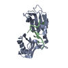
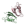
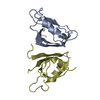
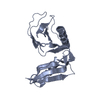
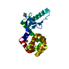
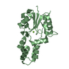
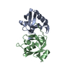
- PDB-1g2e: CRYSTAL STRUCTURE OF HUD AND AU-RICH ELEMENT OF THE TUMOR NECROSI... -
+ Open data

- Basic information
| Entry | Database: PDB / ID: 1g2e | ||||||
|---|---|---|---|---|---|---|---|
| Title | CRYSTAL STRUCTURE OF HUD AND AU-RICH ELEMENT OF THE TUMOR NECROSIS FACTOR ALPHA RNA | ||||||
 Components Components |
| ||||||
 Keywords Keywords | TRANSCRIPTION/RNA / PROTEIN-RNA COMPLEX / HuD / AU-Rich Element / TUMOR NECROSIS FACTOR / TRANSCRIPTION-RNA COMPLEX | ||||||
| Function / homology |  Function and homology information Function and homology informationpositive regulation of 3'-UTR-mediated mRNA stabilization / pre-mRNA intronic pyrimidine-rich binding / protein-RNA adaptor activity / poly(A) binding / mRNA 3'-UTR AU-rich region binding / 3'-UTR-mediated mRNA stabilization / RNA processing / RNA splicing / mRNA 3'-UTR binding / mRNA processing ...positive regulation of 3'-UTR-mediated mRNA stabilization / pre-mRNA intronic pyrimidine-rich binding / protein-RNA adaptor activity / poly(A) binding / mRNA 3'-UTR AU-rich region binding / 3'-UTR-mediated mRNA stabilization / RNA processing / RNA splicing / mRNA 3'-UTR binding / mRNA processing / growth cone / perikaryon / ribonucleoprotein complex / axon / dendrite / cytoplasm Similarity search - Function | ||||||
| Biological species |  Homo sapiens (human) Homo sapiens (human) | ||||||
| Method |  X-RAY DIFFRACTION / MOLECULAR REPLACEMENT / Resolution: 2.3 Å X-RAY DIFFRACTION / MOLECULAR REPLACEMENT / Resolution: 2.3 Å | ||||||
 Authors Authors | Wang, X. / Hall, T.M.T. | ||||||
 Citation Citation | Journal: Nat.Struct.Biol. / Year: 2001 Title: Structural basis for recognition of AU-rich element RNA by the HuD protein. Authors: Wang, X. / Tanaka Hall, T.M. | ||||||
| History |
|
- Structure visualization
| Structure viewer | Molecule: MolmilJmol/JSmol |
|---|
- Downloads & links
-Download
| PDBx/mmCIF format | 1g2e.cif.gz | 49.6 KB | Display | PDBx/mmCIF format |
|---|---|---|---|---|
| PDB format | pdb1g2e.ent.gz | 37.7 KB | Display | PDB format |
| PDBx/mmJSON format | 1g2e.json.gz | Tree view | PDBx/mmJSON format | |
| Others |  Other downloads Other downloads |
-Validation report
| Arichive directory | https://data.pdbj.org/pub/pdb/validation_reports/g2/1g2eftp://data.pdbj.org/pub/pdb/validation_reports/g2/1g2e | HTTPS FTP |
|---|
-Related structure data
| Related structure data |  1fxlSC S: Starting model for refinement C: citing same article ( |
|---|---|
| Similar structure data |


-Links
 PDBj
PDBj- Assembly
| Deposited unit | 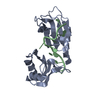
| ||||||||
|---|---|---|---|---|---|---|---|---|---|
| 1 |
| ||||||||
| Unit cell |
|
-Components
| #1: RNA chain | Mass: 3085.820 Da / Num. of mol.: 1 Fragment: FRAGMENT OF AU-RICH ELEMENT OF THE TUMOR NECROSIS FACTOR ALPHA RNA Source method: obtained synthetically |
|---|---|
| #2: Protein | Mass: 18547.186 Da / Num. of mol.: 1 / Fragment: N-TERMINAL TWO RRM-DOMAINS Source method: isolated from a genetically manipulated source Source: (gene. exp.) Homo sapiens (human) / Organ: BRAIN / Plasmid: PPROEXHTC / Species (production host): Escherichia coli / Production host:  |
| #3: Water | ChemComp-HOH /  Mass: 18.015 Da / Num. of mol.: 154 / Source method: isolated from a natural source / Formula: H2O Mass: 18.015 Da / Num. of mol.: 154 / Source method: isolated from a natural source / Formula: H2O |
-Experimental details
-Experiment
| Experiment | Method: X-RAY DIFFRACTION / Number of used crystals: 1 |
|---|
- Sample preparation
| Crystal | Density Matthews: 2.24 Å3/Da / Density % sol: 45.11 % | ||||||||||||||||||||||||||||||
|---|---|---|---|---|---|---|---|---|---|---|---|---|---|---|---|---|---|---|---|---|---|---|---|---|---|---|---|---|---|---|---|
| Crystal grow | Temperature: 293 K / Method: vapor diffusion, hanging drop / pH: 8.5 Details: PEG 3350, Ammonium sulfate, Calcium chloride, Tris, pH 8.5, VAPOR DIFFUSION, HANGING DROP, temperature 293.0K | ||||||||||||||||||||||||||||||
| Components of the solutions |
| ||||||||||||||||||||||||||||||
| Crystal | *PLUS Density % sol: 43 % | ||||||||||||||||||||||||||||||
| Crystal grow | *PLUS Temperature: 20 ℃ | ||||||||||||||||||||||||||||||
| Components of the solutions | *PLUS
|
-Data collection
| Diffraction | Mean temperature: 93 K |
|---|---|
| Diffraction source | Source: ROTATING ANODE / Type: RIGAKU RUH3R / Wavelength: 1.5418 Å |
| Detector | Type: RIGAKU RAXIS IV / Detector: IMAGE PLATE / Date: Apr 5, 2000 |
| Radiation | Protocol: SINGLE WAVELENGTH / Monochromatic (M) / Laue (L): M / Scattering type: x-ray |
| Radiation wavelength | Wavelength: 1.5418 Å / Relative weight: 1 |
| Reflection | Resolution: 2.3→30.38 Å / Num. all: 8327 / Num. obs: 8327 / % possible obs: 97.7 % / Observed criterion σ(I): -3 / Redundancy: 3 % / Biso Wilson estimate: 31.4 Å2 / Rsym value: 0.106 / Net I/σ(I): 8.6 |
| Reflection shell | Resolution: 2.3→2.38 Å / Redundancy: 2.2 % / Mean I/σ(I) obs: 2.1 / Num. unique all: 725 / Rsym value: 0.299 / % possible all: 83.8 |
| Reflection | *PLUS Rmerge(I) obs: 0.106 |
| Reflection shell | *PLUS Highest resolution: 2.3 Å |
- Processing
| Software |
| |||||||||||||||||||||||||
|---|---|---|---|---|---|---|---|---|---|---|---|---|---|---|---|---|---|---|---|---|---|---|---|---|---|---|
| Refinement | Method to determine structure: MOLECULAR REPLACEMENT Starting model: PDB ENTRY 1FXL Resolution: 2.3→30.38 Å / Isotropic thermal model: Isotropic RESTRAINED / Cross valid method: THROUGHOUT / σ(F): 0 / σ(I): 0 / Stereochemistry target values: Engh & Huber Details: A final round of refinement was performed by using all data including reflections set aside for cross-validation in a "free" R-factor set. For last nucleotide A11, only the 5' phosphate ...Details: A final round of refinement was performed by using all data including reflections set aside for cross-validation in a "free" R-factor set. For last nucleotide A11, only the 5' phosphate group and ribose group are observed.
| |||||||||||||||||||||||||
| Displacement parameters | Biso mean: 25.7 Å2 | |||||||||||||||||||||||||
| Refine analyze | Luzzati coordinate error obs: 0.23 Å / Luzzati d res low obs: 5 Å / Luzzati sigma a obs: 0.27 Å | |||||||||||||||||||||||||
| Refinement step | Cycle: LAST / Resolution: 2.3→30.38 Å
| |||||||||||||||||||||||||
| Refine LS restraints |
| |||||||||||||||||||||||||
| Software | *PLUS Name: CNS / Version: 0.9 / Classification: refinement | |||||||||||||||||||||||||
| Refinement | *PLUS Highest resolution: 2.3 Å / σ(F): 0 / % reflection Rfree: 6 % | |||||||||||||||||||||||||
| Solvent computation | *PLUS | |||||||||||||||||||||||||
| Displacement parameters | *PLUS Biso mean: 25.7 Å2 |
