 Movie
Movie Controller

[English] 日本語
 Yorodumi
- PDB-1g1a: THE CRYSTAL STRUCTURE OF DTDP-D-GLUCOSE 4,6-DEHYDRATASE (RMLB)FRO... -
+ Open data

- Basic information
| Entry | Database: PDB / ID: 1g1a | ||||||
|---|---|---|---|---|---|---|---|
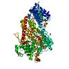
| Title | THE CRYSTAL STRUCTURE OF DTDP-D-GLUCOSE 4,6-DEHYDRATASE (RMLB)FROM SALMONELLA ENTERICA SEROVAR TYPHIMURIUM | ||||||
 Components Components | DTDP-D-GLUCOSE 4,6-DEHYDRATASE | ||||||
 Keywords Keywords | LYASE / Rossmann fold / Protein-NAD complex / Short Chain Dehydrogenase | ||||||
| Function / homology |  Function and homology information Function and homology informationdTDP-glucose 4,6-dehydratase / dTDP-glucose 4,6-dehydratase activity / nucleotide-sugar metabolic process / O antigen biosynthetic process / dTDP-rhamnose biosynthetic process / NADH binding / lipopolysaccharide biosynthetic process / polysaccharide biosynthetic process / nucleotide binding Similarity search - Function | ||||||
| Biological species |  Salmonella enterica subsp. enterica serovar Typhimurium (bacteria) Salmonella enterica subsp. enterica serovar Typhimurium (bacteria) | ||||||
| Method |  X-RAY DIFFRACTION / SYNCHROTRON / Resolution: 2.47 Å X-RAY DIFFRACTION / SYNCHROTRON / Resolution: 2.47 Å | ||||||
 Authors Authors | Allard, S.T.M. / Giraud, M.-F. / Whitfield, C. / Graninger, M. / Messner, P. / Naismith, J.H. | ||||||
 Citation Citation | Journal: J.Mol.Biol. / Year: 2001 Title: The crystal structure of dTDP-D-Glucose 4,6-dehydratase (RmlB) from Salmonella enterica serovar Typhimurium, the second enzyme in the dTDP-l-rhamnose pathway. Authors: Allard, S.T. / Giraud, M.F. / Whitfield, C. / Graninger, M. / Messner, P. / Naismith, J.H. #1: Journal: Acta Crystallogr.,Sect.D / Year: 2000Title: The purifiaction, crystallisation and structural elucidation of dTDP-D-glucose 4,6-dehydratase (RmlB), the second enzyme of the dTDP-L-rhamnose synthesis pathway from Salmonella enterica serovar Typhimurium. Authors: Allard, S.T.M. / Giraud, M.-F. / Whitfield, C. / Messner, P. / Naismith, J.H. | ||||||
| History |
|
- Structure visualization
| Structure viewer | Molecule: MolmilJmol/JSmol |
|---|
- Downloads & links
-Download
| PDBx/mmCIF format | 1g1a.cif.gz | 299.1 KB | Display | PDBx/mmCIF format |
|---|---|---|---|---|
| PDB format | pdb1g1a.ent.gz | 243.9 KB | Display | PDB format |
| PDBx/mmJSON format | 1g1a.json.gz | Tree view | PDBx/mmJSON format | |
| Others |  Other downloads Other downloads |
-Validation report
| Arichive directory | https://data.pdbj.org/pub/pdb/validation_reports/g1/1g1aftp://data.pdbj.org/pub/pdb/validation_reports/g1/1g1a | HTTPS FTP |
|---|
-Related structure data
| Similar structure data |
|---|


-Links
 PDBj
PDBj- Assembly
| Deposited unit | 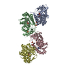
| ||||||||||
|---|---|---|---|---|---|---|---|---|---|---|---|
| 1 | 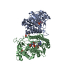
| ||||||||||
| 2 | 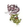
| ||||||||||
| Unit cell |
| ||||||||||
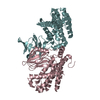
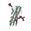
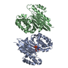
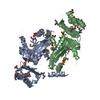
| Details | The biological unit is a dimer. The assymmmetric unit contains two dimers. |
-Components
| #1: Protein | Mass: 40767.516 Da / Num. of mol.: 4 Source method: isolated from a genetically manipulated source Source: (gene. exp.) Salmonella enterica subsp. enterica serovar Typhimurium (bacteria)Species: Salmonella enterica / Strain: subsp. enterica serovar Typhimurium / Gene: RMLB / Variant: SEROVAR TYPHIMURIUM / Plasmid: PET28A(+) / Production host: References: UniProt: P26391, UniProt: Q9EU31*PLUS, dTDP-glucose 4,6-dehydratase #2: Chemical | ChemComp-SO4 /   Mass: 96.063 Da / Num. of mol.: 9 / Source method: obtained synthetically / Formula: SO4 Mass: 96.063 Da / Num. of mol.: 9 / Source method: obtained synthetically / Formula: SO4#3: Chemical | ChemComp-NAD /   Mass: 663.425 Da / Num. of mol.: 4 / Source method: obtained synthetically / Formula: C21H27N7O14P2 / Comment: NAD*YM Mass: 663.425 Da / Num. of mol.: 4 / Source method: obtained synthetically / Formula: C21H27N7O14P2 / Comment: NAD*YM#4: Water | ChemComp-HOH / |  Mass: 18.015 Da / Num. of mol.: 512 / Source method: isolated from a natural source / Formula: H2O Mass: 18.015 Da / Num. of mol.: 512 / Source method: isolated from a natural source / Formula: H2O |
|---|
-Experimental details
-Experiment
| Experiment | Method: X-RAY DIFFRACTION / Number of used crystals: 2 |
|---|
- Sample preparation
| Crystal | Density Matthews: 3.27 Å3/Da / Density % sol: 62.39 % | ||||||||||||||||||||||||||||||
|---|---|---|---|---|---|---|---|---|---|---|---|---|---|---|---|---|---|---|---|---|---|---|---|---|---|---|---|---|---|---|---|
| Crystal grow | Temperature: 293 K / Method: vapor diffusion, sitting drop / pH: 6.3 Details: 0.1M MES, 1.5M lithium Sulfate, pH 6.3, VAPOR DIFFUSION, SITTING DROP, temperature 293K | ||||||||||||||||||||||||||||||
| Crystal grow | *PLUS | ||||||||||||||||||||||||||||||
| Components of the solutions | *PLUS
|
-Data collection
| Diffraction |
| |||||||||||||||
|---|---|---|---|---|---|---|---|---|---|---|---|---|---|---|---|---|
| Diffraction source |
| |||||||||||||||
| Detector |
| |||||||||||||||
| Radiation | Protocol: SINGLE WAVELENGTH / Monochromatic (M) / Laue (L): M / Scattering type: x-ray | |||||||||||||||
| Radiation wavelength |
| |||||||||||||||
| Reflection | Resolution: 2.47→40.8 Å / Num. all: 74855 / Num. obs: 67669 / % possible obs: 90.4 % / Redundancy: 3 % / Biso Wilson estimate: 44.217 Å2 / Rmerge(I) obs: 0.07 / Net I/σ(I): 9.5 | |||||||||||||||
| Reflection shell | Resolution: 2.47→2.56 Å / Redundancy: 2 % / Rmerge(I) obs: 0.358 / Num. unique all: 7903 / % possible all: 72.9 | |||||||||||||||
| Reflection | *PLUS Num. measured all: 201143 | |||||||||||||||
| Reflection shell | *PLUS % possible obs: 72.9 % / Mean I/σ(I) obs: 2.1 |


- Processing
| Software |
| |||||||||||||||||||||||||
|---|---|---|---|---|---|---|---|---|---|---|---|---|---|---|---|---|---|---|---|---|---|---|---|---|---|---|
| Refinement | Resolution: 2.47→40.8 Å / Cross valid method: THROUGHOUT / σ(F): 0 / σ(I): 0 / Stereochemistry target values: Engh & Huber
| |||||||||||||||||||||||||
| Refinement step | Cycle: LAST / Resolution: 2.47→40.8 Å
| |||||||||||||||||||||||||
| Refine LS restraints |
| |||||||||||||||||||||||||
| Software | *PLUS Name: CNS / Classification: refinement | |||||||||||||||||||||||||
| Refine LS restraints | *PLUS
|
