 Movie
Movie Controller

[English] 日本語
 Yorodumi
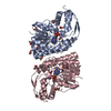
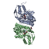
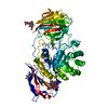
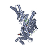
- PDB-4c53: Crystal Structure of Guanarito virus GP2 in the post-fusion confo... -
+ Open data

- Basic information
| Entry | Database: PDB / ID: 4c53 | |||||||||
|---|---|---|---|---|---|---|---|---|---|---|
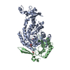
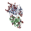
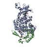
| Title | Crystal Structure of Guanarito virus GP2 in the post-fusion conformation | |||||||||
 Components Components | PRE-GLYCOPROTEIN POLYPROTEIN GP COMPLEX | |||||||||
 Keywords Keywords | VIRAL PROTEIN / ARENAVIRUS / VENEZUELAN HEMORRHAGIC FEVER / GLYCOPROTEIN / FUSION GLYCOPROTEIN | |||||||||
| Function / homology |  Function and homology information Function and homology informationhost cell Golgi membrane / receptor-mediated endocytosis of virus by host cell / host cell endoplasmic reticulum membrane / fusion of virus membrane with host endosome membrane / viral envelope / virion attachment to host cell / host cell plasma membrane / virion membrane / membrane / metal ion binding Similarity search - Function | |||||||||
| Biological species |  GUANARITO VIRUS GUANARITO VIRUS | |||||||||
| Method |  X-RAY DIFFRACTION / SYNCHROTRON / MOLECULAR REPLACEMENT / Resolution: 4.14 Å X-RAY DIFFRACTION / SYNCHROTRON / MOLECULAR REPLACEMENT / Resolution: 4.14 Å | |||||||||
 Authors Authors | Parsy, M. / Huiskonen, J.T. / Harlos, K. / Bowden, T.A. | |||||||||
 Citation Citation | Journal: J.Virol. / Year: 2013 Title: Crystal Structure of Venezuelan Hemorrhagic Fever Virus Fusion Glycoprotein Reveals a Class 1 Post-Fusion Architecture with Extensive Glycosylation. Authors: Parsy, M. / Harlos, K. / Huiskonen, J.T. / Bowden, T.A. | |||||||||
| History |
|
- Structure visualization
| Structure viewer | Molecule: MolmilJmol/JSmol |
|---|
- Downloads & links
-Download
| PDBx/mmCIF format | 4c53.cif.gz | 88.8 KB | Display | PDBx/mmCIF format |
|---|---|---|---|---|
| PDB format | pdb4c53.ent.gz | 69.1 KB | Display | PDB format |
| PDBx/mmJSON format | 4c53.json.gz | Tree view | PDBx/mmJSON format | |
| Others |  Other downloads Other downloads |
-Validation report
| Arichive directory | https://data.pdbj.org/pub/pdb/validation_reports/c5/4c53ftp://data.pdbj.org/pub/pdb/validation_reports/c5/4c53 | HTTPS FTP |
|---|
-Related structure data
| Related structure data |  3mkoS S: Starting model for refinement |
|---|---|
| Similar structure data |

-Links
 PDBj
PDBj- Assembly
| Deposited unit | 
| ||||||||
|---|---|---|---|---|---|---|---|---|---|
| 1 |
| ||||||||
| Unit cell |
|
-Components
| #1: Protein | Mass: 16285.385 Da / Num. of mol.: 3 / Fragment: ECTODOMAIN, RESIDUES 292-418 Source method: isolated from a genetically manipulated source Details: N-LINKED GLYCOSYLATION SEQUONS AT ASN314, ASN351, ASN359, ASN376, AND ASN381 Source: (gene. exp.) GUANARITO VIRUS / Description: SYNTHETICALLY OPTIMIZED CDNA (GENEART) / Plasmid: POPINTTGNEO / Cell line (production host): HUMAN EMBRYONIC KIDNEY 293S / Production host:  HOMO SAPIENS (human) / References: UniProt: A1A3Z2, UniProt: Q8AYW1*PLUS HOMO SAPIENS (human) / References: UniProt: A1A3Z2, UniProt: Q8AYW1*PLUS#2: Polysaccharide | beta-D-mannopyranose-(1-4)-2-acetamido-2-deoxy-beta-D-glucopyranose-(1-4)-2-acetamido-2-deoxy-beta- ...beta-D-mannopyranose-(1-4)-2-acetamido-2-deoxy-beta-D-glucopyranose-(1-4)-2-acetamido-2-deoxy-beta-D-glucopyranose | Source method: isolated from a genetically manipulated source #3: Polysaccharide | 2-acetamido-2-deoxy-beta-D-glucopyranose-(1-4)-2-acetamido-2-deoxy-beta-D-glucopyranose | Source method: isolated from a genetically manipulated source #4: Polysaccharide | Source method: isolated from a genetically manipulated source #5: Sugar |   Type: D-saccharide, beta linking / Mass: 221.208 Da / Num. of mol.: 3 Type: D-saccharide, beta linking / Mass: 221.208 Da / Num. of mol.: 3Source method: isolated from a genetically manipulated source Formula: C8H15NO6 Has protein modification | Y | |
|---|
-Experimental details
-Experiment
| Experiment | Method: X-RAY DIFFRACTION / Number of used crystals: 1 |
|---|
- Sample preparation
| Crystal | Density Matthews: 3.9 Å3/Da / Density % sol: 70 % / Description: NONE |
|---|---|
| Crystal grow | pH: 3.5 Details: 0.15 M LI2SO4, 0.1 M CITRIC ACID PH 3.5, 18% W/V PEG 6000 |
-Data collection
| Diffraction | Mean temperature: 100 K |
|---|---|
| Diffraction source | Source: SYNCHROTRON / Site: Diamond  / Beamline: I04 / Wavelength: 0.953 / Beamline: I04 / Wavelength: 0.953 |
| Detector | Type: ADSC QUANTUM 315 / Detector: CCD / Date: Feb 2, 2013 / Details: MIRRORS |
| Radiation | Monochromator: SINGLE BOUNCE / Protocol: SINGLE WAVELENGTH / Monochromatic (M) / Laue (L): M / Scattering type: x-ray |
| Radiation wavelength | Wavelength: 0.953 Å / Relative weight: 1 |
| Reflection | Resolution: 4.14→50 Å / Num. obs: 5894 / % possible obs: 100 % / Observed criterion σ(I): -3 / Redundancy: 5.1 % / Biso Wilson estimate: 171.92 Å2 / Rmerge(I) obs: 0.09 / Net I/σ(I): 26.6 |
| Reflection shell | Resolution: 4.14→4.14 Å / Redundancy: 5.1 % / Rmerge(I) obs: 1 / Mean I/σ(I) obs: 1.9 / % possible all: 100 |
- Processing
| Software |
| ||||||||||||||||||||||||||||||||||||||||||||||||||||||||||||||||||||||||||||||||||||||||||||||||||||||||||||||||||
|---|---|---|---|---|---|---|---|---|---|---|---|---|---|---|---|---|---|---|---|---|---|---|---|---|---|---|---|---|---|---|---|---|---|---|---|---|---|---|---|---|---|---|---|---|---|---|---|---|---|---|---|---|---|---|---|---|---|---|---|---|---|---|---|---|---|---|---|---|---|---|---|---|---|---|---|---|---|---|---|---|---|---|---|---|---|---|---|---|---|---|---|---|---|---|---|---|---|---|---|---|---|---|---|---|---|---|---|---|---|---|---|---|---|---|---|
| Refinement | Method to determine structure: MOLECULAR REPLACEMENT Starting model: PDB ENTRY 3MKO Resolution: 4.14→38.44 Å / Cor.coef. Fo:Fc: 0.936 / Cor.coef. Fo:Fc free: 0.9076 / Cross valid method: THROUGHOUT / σ(F): 0 / SU Rfree Blow DPI: 0.795
| ||||||||||||||||||||||||||||||||||||||||||||||||||||||||||||||||||||||||||||||||||||||||||||||||||||||||||||||||||
| Displacement parameters | Biso mean: 222.84 Å2
| ||||||||||||||||||||||||||||||||||||||||||||||||||||||||||||||||||||||||||||||||||||||||||||||||||||||||||||||||||
| Refine analyze | Luzzati coordinate error obs: 1.869 Å | ||||||||||||||||||||||||||||||||||||||||||||||||||||||||||||||||||||||||||||||||||||||||||||||||||||||||||||||||||
| Refinement step | Cycle: LAST / Resolution: 4.14→38.44 Å
| ||||||||||||||||||||||||||||||||||||||||||||||||||||||||||||||||||||||||||||||||||||||||||||||||||||||||||||||||||
| Refine LS restraints |
| ||||||||||||||||||||||||||||||||||||||||||||||||||||||||||||||||||||||||||||||||||||||||||||||||||||||||||||||||||
| LS refinement shell | Resolution: 4.14→4.63 Å / Total num. of bins used: 5
|
