HELIX DETERMINATION METHOD: DSSP OUTPUT MODIFIED BY AUTHOR
Remark 700
SHEET DETERMINATION METHOD: DSSP THE SHEETS PRESENTED AS "AD" IN EACH CHAIN ON SHEET RECORDS BELOW ... SHEET DETERMINATION METHOD: DSSP THE SHEETS PRESENTED AS "AD" IN EACH CHAIN ON SHEET RECORDS BELOW IS ACTUALLY AN 8-STRANDED BARREL THIS IS REPRESENTED BY A 9-STRANDED SHEET IN WHICH THE FIRST AND LAST STRANDS ARE IDENTICAL.
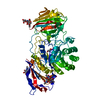
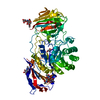
Mass: 74747.906 Da / Num. of mol.: 1 / Fragment: RESIDUES 40-684 Source method: isolated from a genetically manipulated source Details: FRAGMENT CORRESPONDS TO RESIDUES 25-668 BASED ON NUMBERING STARTING AT SECOND UNIPROT INITIATION SITE Source: (gene. exp.) MUS MUSCULUS (house mouse) / Plasmid: PSECTAG2B / Cell line (production host): HEK293 / Production host: HOMO SAPIENS (human) / References: UniProt: P54818, galactosylceramidase
Mass: 18.015 Da / Num. of mol.: 258 / Source method: isolated from a natural source / Formula: H2O
Has protein modification
Y
Nonpolymer details
N-ACETYL-D-GLUCOSAMINE (NAG): THE LAST 3 NUMBERS MATCH THE ASN RESIDUE THEY ARE LINKED TO. THE ...N-ACETYL-D-GLUCOSAMINE (NAG): THE LAST 3 NUMBERS MATCH THE ASN RESIDUE THEY ARE LINKED TO. THE GLYCANS ARE THEN NUMBERED ACCORDING TO THEIR POSITION DISTANT FROM THE ATTACHED ASN.
Sequence details
TWO DIFFERENT START SITES ARE PRESENT IN MOUSE GALC, WE HAVE USED THE SECOND START SITE (TO MATCH ...TWO DIFFERENT START SITES ARE PRESENT IN MOUSE GALC, WE HAVE USED THE SECOND START SITE (TO MATCH THE LITERATURE) WHICH IS M17 IN THE UNIPROT ENTRY. THE FULL CONSTRUCT CONTAINS A SECRETION TAG (THAT IS CLEAVED) A HIS TAG AND A FACTOR XA SITE WHICH ARE NOT.
-
Experimental details
-
Experiment
Experiment
Method: X-RAY DIFFRACTION / Number of used crystals: 1
-
Sample preparation
Crystal
Density Matthews: 3.04 Å3/Da / Density % sol: 59.52 % / Description: NONE
Crystal grow
pH: 6.8 Details: 0.2 M SODIUM ACETATE, 0.1 M SODIUM CACODYLATE, PH 6.8, 34% (W/V) POLYETHYLENE GLYCOL 8000.
Protocol: SINGLE WAVELENGTH / Monochromatic (M) / Laue (L): M / Scattering type: x-ray
Radiation wavelength
Wavelength: 0.98 Å / Relative weight: 1
Reflection
Resolution: 2.1→73.23 Å / Num. obs: 53657 / % possible obs: 99.6 % / Observed criterion σ(I): -3 / Redundancy: 3.7 % / Biso Wilson estimate: 27.18 Å2 / Rmerge(I) obs: 0.12 / Net I/σ(I): 7
Reflection shell
Resolution: 2.1→2.15 Å / Redundancy: 3.7 % / Rmerge(I) obs: 0.7 / Mean I/σ(I) obs: 1.7 / % possible all: 99.8
-
Processing
Software
Name
Version
Classification
PHENIX
(PHENIX.REFINE)
refinement
MOSFLM
datareduction
SCALA
datascaling
PHENIX
phasing
Refinement
Method to determine structure: MIRAS Starting model: NONE Resolution: 2.1→63.182 Å / SU ML: 0.61 / σ(F): 0.88 / Phase error: 20.44 / Stereochemistry target values: ML Details: THE NUMBERS OF REFLECTIONS IN THE TWO TABLES ABOVE (FIT TO DATA USED IN REFINEMENT AND FIT TO DATA USED IN REFINEMENT (IN BINS)) ARE FOR THE DATA BEFORE MERGING OF ANOMALOUS PAIRS.
Rfactor
Num. reflection
% reflection
Rfree
0.2142
5056
5.1 %
Rwork
0.1812
-
-
obs
0.1828
53619
99.45 %
Solvent computation
Shrinkage radii: 0.83 Å / VDW probe radii: 1.1 Å / Solvent model: FLAT BULK SOLVENT MODEL / Bsol: 31.577 Å2 / ksol: 0.331 e/Å3
Displacement parameters
Biso mean: 33.07 Å2
Baniso -1
Baniso -2
Baniso -3
1-
0.9165 Å2
0 Å2
0 Å2
2-
-
0.9165 Å2
0 Å2
3-
-
-
-1.8329 Å2
Refinement step
Cycle: LAST / Resolution: 2.1→63.182 Å
Protein
Nucleic acid
Ligand
Solvent
Total
Num. atoms
5132
0
99
258
5489
Refine LS restraints
Refine-ID
Type
Dev ideal
Number
X-RAY DIFFRACTION
f_bond_d
0.01
5435
X-RAY DIFFRACTION
f_angle_d
1.245
7423
X-RAY DIFFRACTION
f_dihedral_angle_d
16.813
1920
X-RAY DIFFRACTION
f_chiral_restr
0.08
793
X-RAY DIFFRACTION
f_plane_restr
0.006
934
LS refinement shell
Resolution (Å)
Rfactor Rfree
Num. reflection Rfree
Rfactor Rwork
Num. reflection Rwork
Refine-ID
% reflection obs (%)
2.1-2.1239
0.3309
184
0.3011
2983
X-RAY DIFFRACTION
90
2.1239-2.1489
0.3034
140
0.2966
3064
X-RAY DIFFRACTION
91
2.1489-2.1751
0.3003
130
0.2714
3071
X-RAY DIFFRACTION
92
2.1751-2.2026
0.2608
133
0.2693
3075
X-RAY DIFFRACTION
92
2.2026-2.2316
0.2888
164
0.2762
3140
X-RAY DIFFRACTION
92
2.2316-2.2622
0.3121
194
0.2693
3019
X-RAY DIFFRACTION
92
2.2622-2.2945
0.2901
165
0.2562
3115
X-RAY DIFFRACTION
92
2.2945-2.3288
0.2696
164
0.2562
3068
X-RAY DIFFRACTION
93
2.3288-2.3651
0.2872
187
0.2519
3162
X-RAY DIFFRACTION
93
2.3651-2.4039
0.2831
178
0.2444
3071
X-RAY DIFFRACTION
93
2.4039-2.4454
0.2714
196
0.2279
3089
X-RAY DIFFRACTION
93
2.4454-2.4898
0.2815
173
0.2235
3096
X-RAY DIFFRACTION
93
2.4898-2.5377
0.2453
177
0.2196
3131
X-RAY DIFFRACTION
94
2.5377-2.5895
0.2615
172
0.2051
3153
X-RAY DIFFRACTION
94
2.5895-2.6458
0.2167
158
0.1975
3135
X-RAY DIFFRACTION
94
2.6458-2.7074
0.196
185
0.1848
3180
X-RAY DIFFRACTION
95
2.7074-2.7751
0.2214
182
0.1858
3179
X-RAY DIFFRACTION
95
2.7751-2.8501
0.2495
181
0.1839
3140
X-RAY DIFFRACTION
95
2.8501-2.934
0.1896
151
0.1722
3223
X-RAY DIFFRACTION
95
2.934-3.0287
0.2016
165
0.1712
3208
X-RAY DIFFRACTION
96
3.0287-3.1369
0.1964
149
0.1856
3275
X-RAY DIFFRACTION
96
3.1369-3.2625
0.2278
170
0.1826
3207
X-RAY DIFFRACTION
96
3.2625-3.411
0.2007
192
0.1755
3251
X-RAY DIFFRACTION
97
3.411-3.5908
0.208
133
0.1733
3249
X-RAY DIFFRACTION
97
3.5908-3.8158
0.21
179
0.1637
3218
X-RAY DIFFRACTION
96
3.8158-4.1104
0.1947
209
0.1348
3217
X-RAY DIFFRACTION
97
4.1104-4.5239
0.1517
147
0.1253
3222
X-RAY DIFFRACTION
96
4.5239-5.1783
0.1351
167
0.1157
3200
X-RAY DIFFRACTION
96
5.1783-6.523
0.1613
167
0.1501
3134
X-RAY DIFFRACTION
93
6.523-63.2108
0.1767
164
0.1454
3310
X-RAY DIFFRACTION
98
Refinement TLS params.
Method: refined / Refine-ID: X-RAY DIFFRACTION
ID
L11 (°2)
L12 (°2)
L13 (°2)
L22 (°2)
L23 (°2)
L33 (°2)
S11 (Å °)
S12 (Å °)
S13 (Å °)
S21 (Å °)
S22 (Å °)
S23 (Å °)
S31 (Å °)
S32 (Å °)
S33 (Å °)
T11 (Å2)
T12 (Å2)
T13 (Å2)
T22 (Å2)
T23 (Å2)
T33 (Å2)
Origin x (Å)
Origin y (Å)
Origin z (Å)
1
0.8845
-0.0738
-0.2284
0.1439
0.0874
0.314
0.0292
0.141
0.1817
-0.1451
-0.0641
-0.2201
-0.1114
0.129
0.0269
0.3967
-0.1915
0.0887
0.4182
0.0735
0.2877
90.3702
110.2297
1.4877
2
0.4244
0.0289
0.0527
0.3259
-0.023
0.3048
0.0163
0.0169
0.0323
-0.0152
-0.0076
-0.0521
-0.143
0.1813
-0.0037
0.1625
-0.1071
0.009
0.2191
0.0068
0.1191
74.6559
99.4339
22.4809
3
0.5011
0.1702
-0.0489
0.0992
0.0636
0.1605
0.0021
0.0516
0.0572
0.0195
-0.0588
-0.1974
-0.0861
0.113
0.0876
0.2858
-0.2011
0.0056
0.4571
0.0471
0.339
98.4339
105.1553
27.2285
4
1.1741
-0.1353
0.4403
1.3959
-0.2978
0.9063
0.0198
-0.1171
-0.0087
0.1833
-0.0091
-0.1591
-0.0064
0.2286
-0.017
0.1369
-0.0311
-0.0368
0.2726
-0.005
0.1136
82.7605
77.8359
43.6245
Refinement TLS group
ID
Refine-ID
Refine TLS-ID
Selection details
1
X-RAY DIFFRACTION
1
CHAINAAND (RESSEQ25:40ORRESSEQ338:452)
2
X-RAY DIFFRACTION
2
CHAINAANDRESSEQ41:337
3
X-RAY DIFFRACTION
3
CHAINAANDRESSEQ453:471
4
X-RAY DIFFRACTION
4
CHAINAANDRESSEQ472:668
+
About Yorodumi
-
News
-
Feb 9, 2022. New format data for meta-information of EMDB entries
New format data for meta-information of EMDB entries
Version 3 of the EMDB header file is now the official format.
The previous official version 1.9 will be removed from the archive.
In the structure databanks used in Yorodumi, some data are registered as the other names, "COVID-19 virus" and "2019-nCoV". Here are the details of the virus and the list of structure data.
Jan 31, 2019. EMDB accession codes are about to change! (news from PDBe EMDB page)
EMDB accession codes are about to change! (news from PDBe EMDB page)
The allocation of 4 digits for EMDB accession codes will soon come to an end. Whilst these codes will remain in use, new EMDB accession codes will include an additional digit and will expand incrementally as the available range of codes is exhausted. The current 4-digit format prefixed with “EMD-” (i.e. EMD-XXXX) will advance to a 5-digit format (i.e. EMD-XXXXX), and so on. It is currently estimated that the 4-digit codes will be depleted around Spring 2019, at which point the 5-digit format will come into force.
The EM Navigator/Yorodumi systems omit the EMD- prefix.
Related info.:Q: What is EMD? / ID/Accession-code notation in Yorodumi/EM Navigator
Yorodumi is a browser for structure data from EMDB, PDB, SASBDB, etc.
This page is also the successor to EM Navigator detail page, and also detail information page/front-end page for Omokage search.
The word "yorodu" (or yorozu) is an old Japanese word meaning "ten thousand". "mi" (miru) is to see.
Related info.:EMDB / PDB / SASBDB / Comparison of 3 databanks / Yorodumi Search / Aug 31, 2016. New EM Navigator & Yorodumi / Yorodumi Papers / Jmol/JSmol / Function and homology information / Changes in new EM Navigator and Yorodumi
 Movie
Movie Controller


 Open data
 Basic information
 Components
 Keywords
 Function and homology information
 X-RAY DIFFRACTION /
  Authors
 Citation
 Structure visualization
 Downloads & links
 Other downloads


 PDBj
PDBj Assembly
 HOMO SAPIENS (human) / References: UniProt: P54818, galactosylceramidase
 Type: D-saccharide, beta linking / Mass: 221.208 Da / Num. of mol.: 1
 Mass: 40.078 Da / Num. of mol.: 1 / Source method: obtained synthetically / Formula: Ca
 Mass: 18.015 Da / Num. of mol.: 258 / Source method: isolated from a natural source / Formula: H2O
 Sample preparation
 / Beamline: I03 / Wavelength: 0.98
  Processing